Varicose veins are usually explained to patients, and often to ourselves, as a valve problem. The vein wall itself, however, shows reproducible changes in collagen, elastin and smooth muscle, some of them in segments whose valves still work and some of them in the patient's skin. This review sets out what collagen is, which heritable and acquired disorders affect it, how generalised joint hypermobility is assessed, and what the evidence does and does not show about connective-tissue disorders and varicose veins. It pays particular attention to the one disorder in which the diagnosis changes the operation: vascular Ehlers–Danlos syndrome.
The 26-year-old whose veins run in the family
A composite case, typical of referrals to a vein practice
She is 26, has never been pregnant, and has had visible varicose veins on both legs since her late teens. Her mother and her maternal grandmother both had vein surgery before 40. She bruises easily and has sprained each ankle more than once. On examination the forearm skin is soft and velvety and lifts a little further than expected; there are no atrophic scars, no striae and no unusual bruises. Her Beighton score is 6 of 9. Duplex shows bilateral great saphenous reflux from the saphenofemoral junction with incompetent below-knee tributaries, CEAP C2s. When local anaesthetic is infiltrated for a tributary, the needle passes through the skin with almost no resistance. She asks three questions. Is this her connective tissue? Does it change how she should be treated? And will the veins come back?
Collagen in brief
The collagen family comprises 28 members, each containing at least one triple-helical domain built from Gly-X-Y repeats, in which X is often proline and Y often 4-hydroxyproline.1 Glycine at every third position, the imino acids and inter-chain hydrogen bonds stabilise the helix. The fibrillar collagens (types I, II, III, V and XI) form the load-bearing fibrils of skin, tendon, bone and vessel wall; the network-forming type IV builds basement membranes.1
Two fibrillar collagens matter most to the vein practitioner. Type I is the most abundant and in skin forms heterotypic fibrils with type III; type V, co-assembled in small amounts, is thought to regulate fibril diameter.1 Type III is abundant in distensible tissues such as skin and vessel wall, and the proportion of type III within the mixed fibril is widely held to govern how far the tissue can stretch and recover. Collagens I, III and V are all present in the normal and the varicose saphenous wall.2
Two steps of biosynthesis explain the acquired disorders. Prolyl and lysyl hydroxylases need ascorbate, and under-hydroxylated collagen assembles abnormally and is thermally unstable, which is the molecular lesion of scurvy; loss of lysyl hydroxylase 1, encoded by PLOD1, causes kyphoscoliotic Ehlers–Danlos syndrome.3 After secretion, lysyl oxidases begin the cross-links that give mature fibrils their strength.3 Collagen is also long-lived, and advanced glycation end-products accumulate on it with age. Glucosepane, the most prevalent of these cross-links, is roughly 1,500 pmol per mg of skin collagen in people aged 30 to 40 and about 2,700 pmol per mg in diabetes, and it is proposed, although not proven, to stiffen the matrix and to slow its turnover.4
Finally, the mechanism of a mutation decides its severity. A glycine substitution in the helix is incorporated into the trimer and disrupts it; a null allele is cleared by nonsense-mediated decay and leaves half the normal amount of normal protein. In vascular Ehlers–Danlos syndrome, null variants of COL3A1 account for under 5% of recognised variants and delay the onset of complications by almost two decades,5 and functional haploinsufficiency of type V collagen is the working hypothesis for most classical Ehlers–Danlos syndrome.6 Less collagen is milder than wrong collagen.
Heritable and acquired collagen and connective-tissue disorders
The 2017 international classification recognises 13 subtypes of Ehlers–Danlos syndrome (EDS).7 Most are rare and most have a known gene. The important exception is hypermobile EDS (hEDS), which has no identified genetic, epigenetic or metabolomic cause, is diagnosed on clinical criteria alone, and has no molecular test.8 Symptomatic joint hypermobility that does not meet the hEDS criteria is classed as a hypermobility spectrum disorder (HSD). New diagnostic criteria for all EDS types and for HSD have been announced for publication on 1 December 2026 in the American Journal of Medical Genetics; their content is not yet public, existing diagnoses will not need re-evaluation, and an expanded four-joint hypermobility assessment is being tested alongside the Beighton score rather than in place of it.9 The preparatory review found that no single feature or measure completely separates hEDS from HSD.9
Two disorders that overlap clinically with EDS are not collagen disorders at all. Marfan syndrome is caused by FBN1 variants in fibrillin-1, affects about 1 in 5,000 to 10,000, and is diagnosed on an FBN1 variant together with aortic root dilatation or ectopia lentis; it brings striae, hernias and joint laxity, but the GeneReviews chapter does not mention varicose veins.10 Loeys–Dietz syndrome arises in the TGF-β pathway (TGFBR1, TGFBR2, SMAD3, TGFB2, TGFB3 and others), causes aortic dissection at smaller diameters and younger ages than Marfan syndrome, and has velvety, translucent skin, easy bruising and common joint hypermobility; venous disease is not described.11 A third term causes more confusion than either. "Collagen vascular disease" survives from mid-twentieth-century pathology as a label for the autoimmune connective-tissue diseases (systemic lupus erythematosus, systemic sclerosis, dermatomyositis and others). These are not disorders of collagen genes, and this review leaves them aside.
Acquired collagen disorders are more common than any of these and are easy to forget. Ascorbate deficiency disables both hydroxylases.3 Skin collagen falls with age, is lower in women than in men at every age, and dermal thickness tracks collagen content.12 Glycation stiffens long-lived collagen and is accelerated by diabetes.4 Smokers' sun-protected skin shows raised MMP-1 mRNA with no change in TIMP-1, in a small study of 14 smokers and 19 non-smokers.13 And venous hypertension itself produces an acquired fibrosis of the gaiter skin, lipodermatosclerosis, that is the opposite of laxity.
| Disorder | Gene or cause | Key features | Venous relevance |
|---|---|---|---|
| Classical EDS | COL5A1, COL5A2 (AD); functional haploinsufficiency of type V collagen; about 1:20,0006 | Skin hyperextensibility with atrophic scarring; soft, velvety or doughy skin; papyraceous scars; bruising at shins and knees; generalised joint hypermobility6 | Varicose veins in 7 of 122 (6%) in a systematic review; not clearly above background14 |
| Vascular EDS | COL3A1 (AD); about half de novo;15 frequency estimated at 1:50,000 to 1:200,0005 | Arterial rupture, sigmoid perforation, uterine rupture; thin translucent skin with visible veins; bruising at unusual sites; acrogeria; median survival about 50 years15 | Superficial venous insufficiency 41%, mean onset 26 years; early-onset varicose veins a minor criterion; stripping hazardous5,16 |
| Hypermobile EDS | No gene identified; clinical criteria only8 | Generalised joint hypermobility; mild skin hyperextensibility; unusually soft or velvety skin; musculoskeletal complications8 | No controlled, duplex-graded study. GeneReviews lists varicose veins, haemorrhoids and bruising as nonspecific features that do not suggest vascular EDS8 |
| Hypermobility spectrum disorder | No gene; clinical diagnosis9 | Symptomatic joint hypermobility not meeting hEDS criteria | No controlled data; one uncontrolled cohort of standing recruits17 |
| Kyphoscoliotic EDS | PLOD1 (lysyl hydroxylase 1), AR3 | Progressive kyphoscoliosis with joint laxity; defective lysyl hydroxylation | No venous data in the sources reviewed |
| Marfan syndrome | FBN1 (fibrillin-1), AD; 1:5,000–1:10,00010 | Aortic root dilatation, ectopia lentis, skeletal features, striae, hernias, joint laxity10 | Not a collagen disorder; varicose veins not described10 |
| Loeys–Dietz syndrome | TGFBR1/2, SMAD3, TGFB2/3 and others11 | Aortic dissection at small diameters; arterial tortuosity; hypertelorism, bifid uvula; velvety translucent skin; bruising; joint hypermobility common11 | Not a collagen disorder; venous disease not described11 |
| "Collagen vascular disease" | Autoimmune (SLE, systemic sclerosis, dermatomyositis); not collagen genes | Historical misnomer | Outside the scope of this review |
| Scurvy | Ascorbate deficiency; hydroxylase failure3 | Under-hydroxylated, unstable collagen; bruising, bleeding gums, poor wound healing | Capillary fragility; relevant to bruising after procedures |
| Ageing and glycation | Loss of collagen with age; AGE cross-links, higher in diabetes4,12 | Thin, stiff, less extensible skin | Confounds any skin assessment in older patients |
| Lipodermatosclerosis | Venous hypertension, inflammation, fibrosis | Woody induration of the gaiter area (CEAP C4b) | Acquired stiffness; the leg is the wrong place to assess skin laxity |
How common are they?
How often a vein practitioner meets one of these disorders is not known. Population prevalence is about 1 in 20,000 for classical EDS,6 between 1 in 50,000 and 1 in 200,000 for vascular EDS5 and 1 in 5,000 to 10,000 for Marfan syndrome.10 The figure for hypermobile EDS depends on health-record coding: about 1 in 3,100 in a Welsh electronic cohort, as cited by GeneReviews, which describes it as probably an underestimate.8 The prevalence of generalised joint hypermobility itself varies widely with the instrument, the cut-off and the age and sex of the people measured, as the next section explains. No study has measured the prevalence of any heritable connective-tissue disorder among patients presenting with varicose veins, and none of the venous data in this review come from sub-Saharan Africa; the only South African figures in it are the normative joint-mobility data from Phokeng.18
Generalised joint hypermobility and how to assess it
The nine-point score that every vein practitioner should be able to perform was born in South Africa. Beighton, Solomon and Soskolne introduced it in 1973 as a modification of Carter and Wilkinson's manoeuvres, in a census-based survey of 1,081 Tswana people of all ages in Phokeng, north-west of Johannesburg.18 One point is scored for each of: passive dorsiflexion of the little finger beyond 90°, apposition of the thumb to the flexor forearm, hyperextension of the elbow beyond 10°, hyperextension of the knee beyond 10° (each of these on both sides), and placing the palms flat on the floor with the knees straight. Laxity fell rapidly through childhood and more slowly in adults, and females were more mobile than males at every age. Scores of 0 to 2, found in 94% of adult men and 80% of adult women, were regarded as normal for adult Tswana, but no formal cut-off was set.18
The cut-offs in use today are later consensus. The 2017 hEDS criteria define generalised joint hypermobility as a Beighton score of 6 or more before puberty, 5 or more from puberty to age 50, and 4 or more after 50; a score one point below the threshold can be made up by a positive five-point questionnaire.7,8 The five-point questionnaire (5PQ) of Hakim and Grahame asks whether the patient can now, or could ever, place the hands flat on the floor with the knees straight; bend the thumb to the forearm; do the splits or contortions as a child; has dislocated a shoulder or kneecap more than once; and considers themselves double-jointed. Two or more "yes" answers are positive, with sensitivity 84% and specificity 89% in the derivation cohort and 84% and 80% in a second cohort.19 It captures historical hypermobility in adults whose Beighton score has fallen with age.8
The score has limits. It was built for epidemiological screening, not diagnosis; its joints are predominantly in the upper limb, it omits many major joints, and it should not be used alone to rule generalised joint hypermobility in or out.20 Reported prevalence varies widely with the instrument, the cut-off and the age and sex of the population, so no single figure for "how common" is meaningful without those details.18,20
Skin should be examined at the same time, at the volar forearm of the non-dominant arm, lifted between finger and thumb. The numerical thresholds for hyperextensibility (more than 1.5 cm at the distal forearm and dorsum of the hand, more than 3 cm at the neck, elbows and knees) belong to classical EDS and derive from Remvig's work; the hEDS criteria specify only "mild skin hyperextensibility" and "unusually soft or velvety skin".6,8,21 Instrumented measurement has been tried in very small numbers (6 patients with EDS, 11 with joint hypermobility syndrome, 19 controls): suction extensibility did not reach significance, a stiffness meter did, and inter-examiner agreement for the clinical tests ranged from 0.44 to 0.72.21 High-frequency ultrasound in 21 patients with classical and 9 with hypermobile EDS found the skin significantly thinner than in controls, most markedly at the chest and the distal lower leg.22 That last site is exactly where chronic venous disease thickens the skin, so in a vein patient any skin assessment belongs on the forearm or trunk, not the leg.
The varicose vein wall
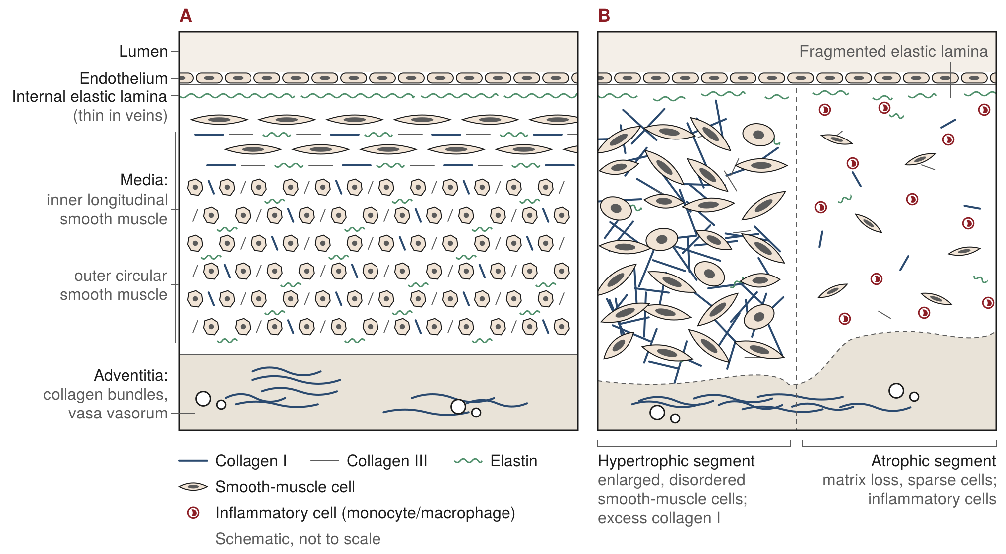
The saphenous wall has three layers: an intima of endothelium on a basement membrane with an internal elastic lamina; a media with inner longitudinal and outer circular smooth-muscle layers set in collagen and elastin; and an adventitia carrying the vasa vasorum.23 In varicose veins the clear boundaries between layers are lost, elastin is fragmented, and smooth-muscle cells are disorganised, dedifferentiated and secrete more MMP-2.23 The wall is not simply thinned. It is a patchwork of hypertrophic segments, with enlarged smooth-muscle cells and excess matrix, and atrophic segments, with matrix degradation and inflammatory infiltrate.23
Total collagen. The findings conflict. Gandhi and colleagues found more collagen (189 vs 144 mg/g), less elastin (53 vs 74 mg/g) and a collagen-to-elastin ratio of 3.7 against 2.0 in normal vein.24 Psaila and Melhuish found the opposite: less collagen (51 vs 70 µg/mg), together with lower breaking strength and greater stiffness.25 Others found no change, and the 2021 review by Raffetto and Khalil lists collagen content as increased, decreased or unchanged depending on the study.23 Method, segment and disease stage probably explain much of this; total collagen is not the finding to build on.
Collagen I and III. The more reproducible observation is a shift in the ratio. Waksman and colleagues compared 21 normal veins with veins from 37 varicose subjects: collagen I was higher (p<0.01) in both the affected and the unaffected segments, collagen III was slightly lower, and the ratio of type I to types III and V rose (p<0.02), which the authors read as support for the "weak wall" theory.2 The Servier group then took the question into culture. In smooth-muscle cells grown from varicose veins, collagen I was overproduced with increased mRNA and collagen III was reduced.26 In a separate study of dermal fibroblasts from patients with varicose veins, collagen I synthesis and mRNA were again raised, collagen III synthesis was reduced with its mRNA unchanged, and of the metalloproteinases and inhibitors assayed only proMMP-2 was raised; the authors concluded that the dysregulation is present in skin as well as vein, and is therefore likely to be generalised rather than confined to the vein.27 The collagen III deficit was then traced to degradation rather than synthesis: mRNA, mRNA stability and translation were normal, the protein was degraded faster, MMP-3 was raised, and the MMP inhibitor marimastat partly restored production.28 The group proposed that loss of type III, which confers elasticity, accounts for the abnormal distensibility of the varicose wall.26,27
These are small in vitro studies from one laboratory and have not been independently replicated in the same form. Two pieces of evidence sit either side of them. The same investigator later reported at a congress workshop that groin skin biopsies from patients with venous ulcers showed a similar pattern, but noted that sampling the groin, close to diseased veins, could partly account for the result; the finding therefore does not settle whether the change is systemic.29 As support, an independent proteomic study of six varicose and six normal saphenous veins found collagen I α1 and α2 chains raised about 2.5-fold, collagen III α1 reduced to a quarter (p=0.043), loss of the small leucine-rich proteoglycans, and TIMP-3 protein reduced to 5% of control with its transcript unchanged.30 Different method, same direction.
MMPs, TIMPs and inflammation. Metalloproteinase data also conflict, and here the conflict is probably real rather than methodological. Some groups found high TIMP-1 with low MMP-2, a balance that favours matrix accumulation; others found raised MMP-1, -2, -3 and -9, a balance that favours degradation; the 2021 review lists these results as contradictory.23 Both may be true in different segments of the same vein. What is not disputed is inflammation: in 17 saphenous veins removed at venous surgery, monocytes and macrophages infiltrated the valve leaflets and wall in every stained specimen, heaviest in the valve sinus, with collagen degeneration in all specimens examined and none of these changes in control veins.31
Wall first, or valve first? The valve-first account describes descending reflux from the junction, with leucocyte-damaged, shortened leaflets as its histological signature; the monocyte infiltration of the valve sinus fits it well.31 The wall-first account rests on three observations. First, matrix abnormalities are present in still-competent segments: in Gandhi's series, duplex-competent great saphenous segments from limbs with varices elsewhere had a collagen-to-elastin ratio of 3.9, the same as the varicose segments, although only four such segments were studied, so this suggests rather than shows.24 Waksman's unaffected segments carried the same collagen I excess as the affected ones.2 Second, the dermal fibroblast findings, if confirmed, place the defect outside the vein altogether.27 Third, duplex epidemiology does not match a purely descending process. In subjects aged 15 to 35 with superficial incompetence, Labropoulos and colleagues found the below-knee great saphenous vein involved in 68% of limbs and the saphenofemoral junction in only 32%, with multisegmental reflux in 95% of varicose limbs; they concluded that reflux appears to be a local or multifocal process in addition to, or separate from, a retrograde one.32
The fair reading is that both are true and that neither camp is settled. Wall and valve abnormalities coexist, venous hypertension feeds back through inflammation and metalloproteinases on both, and the sequence probably differs between patients and between segments. The wall is part of the disease. It is not the whole story, and the evidence that it comes first is suggestive, not conclusive.
What genetics adds
Varicose veins are familial and moderately heritable. In 4,033 German nuclear families covering 16,434 people, narrow-sense heritability was 17.3% (SE 2.5%), with age and sex explaining a further 10.7% of risk.33 In the Tampere study of 6,874 people aged 40 to 60, a family history carried an odds ratio of 4.9, female sex 2.2, number of births 1.2 to 2.8, standing work 1.6 and height 1.4.34
Genome-wide association studies have now reached about 135,000 cases. In the UK Biobank, Fukaya and colleagues confirmed height as an independent risk factor, with a hazard ratio of 1.74 (95% CI 1.51 to 2.01) for the top against the bottom quartile and support from Mendelian randomisation, and found 30 new loci including CASZ1, PIEZO1 and PPP3R1; FBN2 appeared as an expression signal.35 Ahmed and colleagues, in 135,514 cases and 675,111 controls of European ancestry, reported 49 signals at 46 replicated loci, SNP heritability of 5 to 8%, and matrix genes among them (EFEMP1, encoding fibulin-3, and COL27A1); the Mendelian-randomisation effect of hernia on varicose veins was described as suggestive, and a polygenic score was higher in those with a history of surgery, an association rather than a demonstrated prediction.36 Levin and colleagues, in a multi-ancestry analysis of 49,765 cases, found 139 loci; the cohort included about 2,500 participants of African ancestry, but ancestry-specific results are not reported in the main text, and no African-ancestry-specific findings have been published.37 That gap matters in the country where the Beighton score was first applied.
Two conclusions follow. The classic EDS collagen genes and FBN1 are not among the reported lead loci in any of these studies, although this has not been checked exhaustively against the full locus tables.35–37 Common varicose veins are therefore not a mild form of a monogenic collagen disorder. And the strongest signals point to vascular development, mechanosensation and valve biology rather than to collagen structure.35,36 In lymphoedema–distichiasis, FOXC2 variants were associated with great saphenous reflux in all 18 carriers, including three without lymphoedema, against 1 of 12 referents, and the authors called FOXC2 the first gene strongly associated with primary venous valve failure.38 The genetics, in short, supports both sides of the wall–valve debate: a polygenic matrix contribution and a polygenic valve contribution. A plain-language summary for patients is at varicose veins and genetics.
Connective-tissue disorders and venous disease: the clinical evidence
It is often stated that varicose veins are strikingly more prevalent across the Ehlers–Danlos syndromes. The evidence does not support the generalisation. Only vascular EDS has quantified venous data, and in the other types the rate is not clearly above background.
| Condition or marker | Venous finding | Source and strength |
|---|---|---|
| Vascular EDS | Superficial venous insufficiency in 41% of 68 COL3A1 patients (37% symptomatic, 4% asymptomatic) against 17–23% in an external reference population (p=0.008); mean onset 26 years; 44% before age 20; mostly C2–C3, none C5–C6. Procedural outcomes in Table 3 | Frank 2015, French reference centre; unmatched external comparator; small treatment numbers16 |
| Vascular EDS | Early-onset varicose veins (before 30, not pregnancy-related) listed as a clue prompting COL3A1 testing; present in 15% of the Sheffield cohort | Bowen 2023, UK national diagnostic service; denominator not stated39 |
| Classical EDS | Varicose veins in 7 of 122 (6%): COL5A1 4 of 110, COL1A1 3 of 12 | D'hondt 2018, systematic review of nonvascular EDS types; hypermobile EDS excluded14 |
| Hypermobile EDS / HSD | No controlled, duplex-graded CEAP study found | — |
| Generalised joint hypermobility | 718 male Iranian recruits, 211 (29.3%) hypermobile (mean Beighton 5.5 vs 1.2). More spider and varicose veins at baseline; venous insufficiency rose after three months of standing duty only in the hypermobile group (non-hypermobile p=0.25) | Azma 2015; clinical CEAP grading only, no duplex, no effect sizes, hypermobility cut-off not stated, men only17 |
| Ankle dorsiflexion | Each 5° of additional dorsiflexion: OR 1.11 for progression of chronic venous disease over 11 years (p=0.01); laxity offered as the explanation | Kim 2015, San Diego Population Study, 1,025 people40 |
| Hernia | Shared locus (EFEMP1); Mendelian-randomisation effect of hernia on varicose veins "suggestive"; no clinical odds ratio found | Ahmed 2022, GWAS36 |
| Pregnancy and parity | Number of births OR 1.2–2.8 (adjusted); female sex OR 2.2 | Laurikka 2002, population study34 |
| Marfan syndrome | No prevalence data; GeneReviews does not mention varicose veins | —10 |
| Other EDS, endovenous treatment | 1940-nm laser plus foam in spondylodysplastic and classical EDS: occluded at one year, no peri-procedural complications, exaggerated sclerosant reactions managed conservatively | Braß 2025, two case reports41 |
Three points stand out. First, the vascular EDS data are the only data of any quality, and they show a disease that is roughly twice as common as background, starts decades early, stays at low CEAP class, and is dangerous to operate on through the groin. Second, in hypermobile EDS and HSD, which together account for most hypermobile patients a vein practitioner will meet, there is no controlled study at all; the Azma cohort is the only prospective test of hypermobility against a venous outcome, and it used clinical grading in young men without duplex.17 Third, the "connective-tissue weakness" link with hernia is real at the genetic level but modest, and nothing in it has been shown to predict the outcome of treatment.36
Pregnancy deserves a separate word, because it is the connective-tissue argument most often made to patients. Parity raises risk in a dose-related way,34 and hormonal, volume and pressure effects all contribute. Relaxin is regularly invoked as the link between pregnancy, laxity and veins. No study of relaxin and the human vein wall could be found for this review, and the claim should not be made.
Soft skin, tough skin
Any practitioner who infiltrates local anaesthetic several hundred times a year notices that skin varies. In some patients the needle passes with almost no resistance; in others it has to be pushed. It is tempting to read the first as lax connective tissue and the second as robust connective tissue, and to wonder whether either predicts how the veins will behave. What can be inferred, and what cannot?
Soft, velvety or hyperextensible skin is a recognised feature of classical and hypermobile EDS and of Loeys–Dietz syndrome, and thin, translucent skin with visible veins is a minor criterion of vascular EDS.6–8,11 Skin is thinner on ultrasound in both classical and hypermobile EDS.22 So a young patient whose forearm skin is unusually soft and lifts further than expected has a finding that is worth a Beighton score, a five-point questionnaire and a family history.
Tough skin is harder to interpret, because it has several causes that have nothing to do with collagen genes. Skin collagen is lower in women than in men at every age, so male skin is thicker.12 Collagen is lost with age, and what remains has accumulated glycation cross-links that stiffen it, more so in diabetes.4,12 Smoking raises MMP-1 expression in skin,13 and sun exposure remodels the dermis. In the leg of a vein patient, lipodermatosclerosis adds acquired fibrosis on top of all of this. Tough skin, in other words, tells the practitioner about age, sex, sun, smoking, glycaemia and venous disease. It does not tell them that the collagen is sound. Nor does tough mean strong: the varicose vein wall of Psaila and Melhuish was stiffer than normal and had lower breaking strength at the same time.25
The limit is this. No study has measured needle-puncture force in living skin against collagen content, skin extensibility or hypermobility. The observation is a clinical impression without a validated measure behind it, and it should be recorded as such. If it is to become more than that, it would need a standardised site away from the leg, an instrumented measure, and blinded comparison with the Beighton score and skin extensibility, none of which has been done.
Vascular EDS: what the vein practitioner must not miss
Vascular EDS is rare, with a frequency estimated at between 1 in 50,000 and 1 in 200,000,5 and about half of cases arise de novo, so a negative family history does not exclude it.15 Median survival is about 50 years, about five years shorter in men than in women; arterial rupture, most often in the thorax or abdomen, is the characteristic complication; and maternal mortality is about 5% per pregnancy.15 In the series of Pepin and colleagues, 220 index patients with biochemically confirmed disease and 199 affected relatives, complications were rare in childhood, about a quarter of index patients had a first complication by age 20 and more than 80% by age 40, and the calculated median survival was 48 years; most deaths were from arterial rupture.42 Diagnosis rests on COL3A1 testing. The 2017 criteria list "early-onset varicose veins (under age 30 and nulliparous if female)" among the minor criteria, alongside thin translucent skin with increased venous visibility, bruising unrelated to trauma or at unusual sites, acrogeria, spontaneous pneumothorax and small-joint hypermobility; the major criteria are a family history with a documented COL3A1 variant, arterial rupture at a young age, spontaneous sigmoid perforation, third-trimester uterine rupture and carotid-cavernous fistula.7
The venous facts (Table 2) come from a single French cohort of 68 patients with confirmed COL3A1 variants, in whom superficial venous insufficiency was present in 41%, began on average at 26 years, and was mostly C2 to C3.16 Of nine who had undergone saphenous stripping, two had serious vascular complications, one a femoral vein rupture in an 18-year-old and one haemorrhagic shock at crossectomy, and all nine recurred; five treated with foam and one with radiofrequency ablation had no vascular complications.16 The international consensus that followed states that early-onset varicose veins should raise the suspicion of vascular EDS, that stripping should be avoided because of the risk of extensive vascular damage and uncontrolled bleeding, that compression is the primary therapy, that endovenous laser and radiofrequency ablation have been reported successfully and that foam may be a complementary or alternative treatment; it adds that an unusually complicated stripping can itself be the event that prompts the diagnosis.5 GeneReviews adds that elective surgery is generally discouraged unless the benefit is expected to be substantial and that conventional arteriography should be discouraged.15 The UK national diagnostic service lists early-onset varicose veins, before 30 and unrelated to pregnancy, among the clues that prompt testing.39
Table 3 sets out the rates behind these statements. The venous figures all come from one cohort, and the procedural figures from nine stripped and six endovenously treated patients; no other series was found for this review.
| Complication | Reported rate | Population/source |
|---|---|---|
| Any major complication (arterial, bowel or uterine rupture) | About 25% of index patients by age 20; more than 80% by age 40; median survival 48 years | 220 index patients with biochemically confirmed vascular EDS and 199 affected relatives, Pepin 200042 |
| Arterial rupture | The commonest cause of death; sites thorax and abdomen 66%, head and neck 17%, limbs 17% | Pepin 2000 (cause of death);42 GeneReviews (sites)15 |
| Bowel rupture, usually sigmoid | About 25–30% over a lifetime; rarely fatal | International consensus 20175 |
| Spontaneous pneumothorax | 12%, often the first manifestation | International consensus 20175 |
| Pregnancy-related death | About 5% of affected women, half of them in a first pregnancy; in the series of Pepin and colleagues 12 of 81 women who became pregnant died of pregnancy complications | International consensus 2017;5 Pepin 200042 |
| Superficial venous insufficiency | 41% (37% symptomatic, 4% asymptomatic) against 17–23% in an external reference population, p=0.008; mean onset 26 years; 44% before age 20; mostly C2–C3 | 68 patients with confirmed COL3A1 variants, French reference centre16 |
| Serious vascular complication of saphenous stripping or crossectomy | 2 of 9 (one femoral vein rupture in an 18-year-old; one haemorrhagic shock at crossectomy) | Same cohort16 |
| Recurrence after stripping | 9 of 9 (8 symptomatic) | Same cohort16 |
| Vascular complication after ultrasound-guided foam (5) or radiofrequency ablation (1) | 0 of 6, early or late | Same cohort; treated after compression had failed16 |
| Peri-procedural complication of 1940-nm endovenous laser with foam, non-vascular EDS | 0 of 2; exaggerated inflammatory reactions to the sclerosant reported and managed conservatively; both trunks occluded at one year | One spondylodysplastic and one classical EDS patient, case reports41 |
| Varicose veins in classical EDS (for comparison) | 7 of 122 (6%) | Systematic review of nonvascular EDS types; hypermobile EDS excluded14 |
| Recurrent varices after endovenous great saphenous ablation, general population (for comparison) | 22% of limbs at two years or more; recanalisation accounts for 32% of recurrences | Systematic review of randomised trials43 |
Before you treat: red flags for vascular EDS in a varicose-vein referral
- Varicose veins beginning before 30, not explained by pregnancy7,39
- Thin, translucent skin with increased venous visibility7
- Bruising without trauma, or at unusual sites such as the cheeks or back7
- Acrogeria: a prematurely aged appearance of the hands and feet7
- Personal or family history of arterial rupture, dissection or aneurysm at a young age, of spontaneous bowel perforation, or of third-trimester uterine rupture7,15
- Spontaneous pneumothorax, carotid-cavernous fistula, tendon or muscle rupture7
- A previous vein operation that bled or ruptured out of proportion to the procedure5
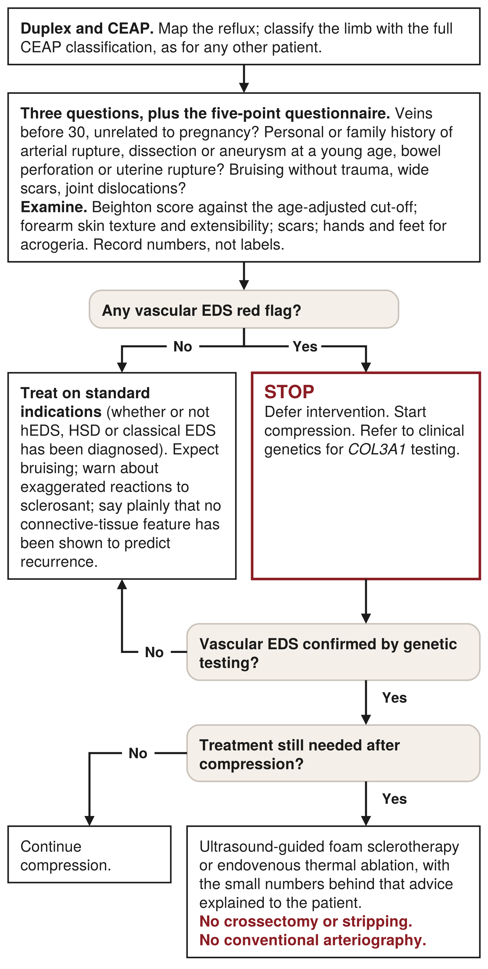
If any of these is present: defer intervention, refer to clinical genetics for COL3A1 testing, and start compression. If treatment is still needed once the diagnosis is known, avoid crossectomy and stripping; ultrasound-guided foam sclerotherapy and endovenous thermal ablation have been reported without vascular complications in small numbers.5,16 Avoid conventional arteriography.15
The corollary is equally important. The GeneReviews chapter on hypermobile EDS is explicit that nonspecific venous and haematological features, including varicose veins, haemorrhoids and easy bruising, are not suggestive of vascular EDS on their own.8 The 26-year-old in the case above has early-onset bilateral varicose veins, a minor criterion, and easy bruising of the nonspecific kind; she has no thin or translucent skin, no bruising at unusual sites, no acrogeria and no family history of rupture. She warrants a careful history for rupture events in the family and a look at her skin, hands and feet, not automatic genetic testing. The practitioner who tests every young patient with varicose veins will find very few cases and alarm many families; the practitioner who never asks will one day strip a vascular EDS patient.
What the guidelines say
Guideline content on heritable connective-tissue disorders
ESVS 2022. In the sections reviewed for this article (the introduction, epidemiology and pathophysiology chapters), the European guideline does not mention Ehlers–Danlos syndrome, Marfan syndrome, connective-tissue disorders, hypermobility or metalloproteinases. It lists a positive family history and parity among risk factors and describes degenerative loss of elastin and collagen with fibrosis in the vein wall.44 Later chapters were not reviewed, so specific content there cannot be excluded.
Vascular EDS consensus 2017 and GeneReviews. These are the only documents found with explicit venous advice: compression first; avoid stripping; endovenous ablation and foam reported as successful or acceptable alternatives; elective surgery generally discouraged.5,15
The European venous guideline, in other words, does not tell the practitioner what to do with a hypermobile patient, because there is no evidence on which to base a recommendation, and no guidance specific to heritable connective-tissue disorders was found in the North American guideline summary either. Venous management in vascular EDS rests on a consensus statement and a handful of treated cases. No drug has been shown to alter vein-wall collagen or metalloproteinase activity in humans; the marimastat effect on collagen III was observed in cell culture and is not a treatment.28
Management: practical implications
What follows is the author's synthesis, not a guideline.
History. Three questions take a minute and change management. At what age did the veins appear, and was it before 30 and unrelated to pregnancy? Is there a family history not only of vein surgery but of arterial rupture, aneurysm or dissection at a young age, bowel perforation or uterine rupture? Does the patient bruise without trauma, heal with wide scars, or dislocate joints? The five-point questionnaire adds thirty seconds. Height, parity and family history of varicose veins are established risk factors and belong in the record.34,35
Examination. A Beighton score, with the age-adjusted 2017 cut-off, and a look at the forearm skin and any scars. Record the score as a number, not as "hypermobile", because the cut-off may change with the 2026 criteria. Classify the limb with the full CEAP classification, because the vascular EDS literature is expressed in those terms and because low CEAP class in a young patient is itself informative.
Treatment choice. In classical EDS, hypermobile EDS and HSD there is no evidence of a higher rate of varicose veins and no evidence of worse outcomes; treat on standard indications. Be alert to bruising and exaggerated sclerosant reactions, which are reported at case level.41 Tumescent radiofrequency ablation, ultrasound-guided foam sclerotherapy and medical adhesive closure all avoid the groin dissection that is the hazard in vascular EDS. Foam and radiofrequency ablation have been reported in six vascular EDS patients in all; no report of medical adhesive closure in any heritable connective-tissue disorder was found for this review. In suspected or confirmed vascular EDS, follow the consensus: compression, genetics, and no stripping. Table 4 summarises the options and the evidence behind each.
| Option | Best evidence | Practical points |
|---|---|---|
| Compression | Primary therapy for superficial venous insufficiency in vascular EDS in the 2017 international consensus;5 no trial data in any heritable connective-tissue disorder | The first measure while a suspected vascular EDS is investigated; the six endovenously treated patients in the French cohort had first failed compression16 |
| Ultrasound-guided foam sclerotherapy | Five vascular EDS patients without early or late vascular complications;16 "complementary or alternative" in the consensus;5 exaggerated inflammatory reactions to foam reported in non-vascular EDS case reports41 | No groin dissection and no tumescent infiltration; warn about bruising and exaggerated local reactions; the numbers are too small to quote an occlusion or recurrence rate |
| Radiofrequency ablation | One vascular EDS patient without complications;16 listed with endovenous laser as "successfully reported" in the consensus5 | Percutaneous, no groin incision; no occlusion or recurrence data in vascular EDS; no comparison between thermal modalities in any EDS type |
| Endovenous laser ablation | Reported successfully in vascular EDS according to the consensus;5 1940-nm laser with foam occluded at one year in one spondylodysplastic and one classical EDS patient41 | Same considerations as radiofrequency ablation; the two published cases had no intra- or postoperative complications |
| Medical adhesive closure | No report in any heritable connective-tissue disorder was found for this review | Avoids tumescent anaesthesia and thermal energy; the absence of reports is an absence of data, not reassurance |
| Crossectomy and stripping | Serious vascular complications in 2 of 9 and recurrence in 9 of 9 vascular EDS patients;16 "should be avoided" in the consensus5 | Not to be performed in suspected or confirmed vascular EDS; an unusually complicated stripping may itself be the event that prompts the diagnosis5 |
| Standard indications (classical EDS, hypermobile EDS, HSD) | No evidence of a higher prevalence of varicose veins14 or of worse outcomes; case reports only41 | Treat as any other patient; record the Beighton score as a number; expect bruising; counsel about recurrence from the general figures43 |
Counselling about recurrence. Patients with hypermobility often ask whether their veins are more likely to come back. The answer should be given plainly: it is not known. In randomised trials with at least two years of follow-up, 22% of limbs had recurrent varices after endovenous ablation of the great saphenous vein, the same proportion as after ligation and stripping. Among the 125 recurrent limbs after ablation, recanalisation accounted for 32%, anterior accessory saphenous incompetence for 19%, calf perforators for 7% and neovascularisation for 2%; after stripping, neovascularisation accounted for 18%.43 Recanalisation of the treated trunk is therefore the single largest cause of recurrence after ablation, and it is a technical and anatomical event rather than a connective-tissue one. No study has tested hypermobility, skin laxity or any other connective-tissue phenotype as a predictor of recurrence after any varicose-vein treatment. The only recurrence data in a heritable connective-tissue disorder are the nine stripped vascular EDS patients, all of whom recurred.16 A biologically plausible hypothesis is not a prognosis, and it should not be offered to patients as one. A plain-language account of the same evidence, written for patients, is at collagen, connective tissue and varicose veins.
Documentation. A practitioner who records a Beighton score, or the needle impression, in every patient should record what was measured and how, and should not use it to counsel until it has been tested.
A suggested approach to the hypermobile or soft-skinned patient with varicose veins (the author's synthesis, not a guideline)
- Map the reflux with duplex and classify the limb with the full CEAP classification, as for any other patient.
- Ask three questions: did the veins appear before 30, unrelated to pregnancy; is there a personal or family history of arterial rupture, dissection or aneurysm at a young age, bowel perforation or uterine rupture; does the patient bruise without trauma, heal with wide scars or dislocate joints. Add the five-point questionnaire.7,19,39
- Examine: Beighton score against the age-adjusted cut-off, forearm skin texture and extensibility, scars, and the hands and feet for acrogeria. Record numbers, not labels.7,8
- Any vascular EDS red flag (box above): defer intervention, start compression and refer to clinical genetics for COL3A1 testing.5,15,39
- Hypermobility or soft skin without a red flag, or red flags with vascular EDS not confirmed on genetic testing, whether or not a diagnosis of hypermobile EDS, HSD or classical EDS has been made: treat on standard indications; expect bruising; warn about exaggerated reactions to sclerosant; say plainly that no connective-tissue feature has been shown to predict recurrence.14,41,43
- Confirmed vascular EDS still needing treatment after compression: ultrasound-guided foam or endovenous thermal ablation, with the small numbers behind that advice explained to the patient; no crossectomy or stripping; no conventional arteriography.5,15,16
In the author's practice
Dr Blignaut's practice offers radiofrequency ablation, medical adhesive (cyanoacrylate) closure and ultrasound-guided foam sclerotherapy at three centres, in Fourways, Umhlanga and Somerset West; it does not offer endovenous laser or mechanochemical ablation. Every patient has a duplex scan before treatment. About 750 patients a year undergo radiofrequency ablation across the three centres.
His observation, which prompted this review, is that at the needle, whether for local anaesthetic infiltration or for access punctures, some patients' skin is very soft and is pierced with almost no resistance, while others' is very tough. This is an informal clinical impression. It has not been recorded systematically, it is not a validated measure, and it has not been compared with any assessment of hypermobility, skin extensibility or treatment outcome. It is reported here as an unaudited clinical observation and as a question, not as a finding.
Areas of uncertainty
- Is the varicose wall abnormal before the valve fails? The competent-segment data rest on four segments in one series and on unaffected segments in another; the dermal-fibroblast finding has not been independently replicated in the same form.2,24,27
- Are varicose veins more common in hypermobile EDS or HSD? No controlled, duplex-graded study exists.
- Does any connective-tissue phenotype predict recurrence after treatment? Unknown; no study has tested it.
- What is the MMP/TIMP balance in a given segment, and does it matter for treatment? Reports conflict, and no drug has been shown to change it in humans.23
- What does the needle feel? No study has measured puncture force in living skin against collagen, extensibility or hypermobility.
- Do the genetic findings apply to African populations? No ancestry-specific results have been published.37
- What will the December 2026 EDS/HSD criteria change? The content is not yet public.9
- How large is the venous burden in vascular EDS outside France? The 41% figure comes from one cohort with an external comparator.16
Key clinical points
- The varicose vein wall is part of the disease. It shows a reproducible shift towards collagen I and away from collagen III, fragmented elastin and disorganised smooth muscle, some of it in segments that are still competent.2,24,30
- Wall and valve both contribute. The evidence for a wall-first sequence is suggestive, not conclusive; the two feed each other through venous hypertension and inflammation.
- Common varicose veins are polygenic, with heritability of about 17%, and the lead loci point to vascular development and mechanosensation; the EDS collagen genes are not among them.33,35–37
- Only vascular EDS has quantified venous data: superficial venous insufficiency in 41%, mean onset 26 years, mostly C2–C3.16
- In vascular EDS, do not strip. Two of nine stripped patients had serious vascular complications and all nine recurred; compression is first-line, and foam or endovenous ablation have been used without complications in small numbers.5,16
- Varicose veins before 30, not pregnancy-related, are a minor criterion for vascular EDS and a recognised prompt for COL3A1 testing when other features are present.7,39
- Hypermobile EDS has no gene and no controlled venous data. Varicose veins, haemorrhoids and bruising on their own do not suggest vascular EDS.8
- Score hypermobility properly: Beighton with the 2017 age-adjusted cut-offs, the five-point questionnaire for historical laxity, and skin examined at the forearm, never the leg.7,19,20
- Tough skin is not strong skin. Age, male sex, glycation, sun, smoking and lipodermatosclerosis all stiffen it; no study links needle resistance to collagen or hypermobility.
- Tell patients the truth about recurrence: about one limb in five at two years or more, with recanalisation the largest single cause, and no evidence that hypermobility changes that figure.43
A bedside rule
Varicose veins before 30 + not explained by pregnancy + thin or translucent skin, unusual bruising or a family history of rupture = ask about vascular EDS, refer for COL3A1 testing, and do not operate through the groin until you know.
Disclosure. The author performs endovenous and other venous procedures in private practice. This article is for medical education and does not replace individual clinical assessment.
Figures. Diagrams drawn for this review by Vein Centres South Africa; clinical photographs as credited.
How to cite
Blignaut J. The wall, not the valve: collagen, connective tissue disorders and varicose veins. Clinical review. Vein Centres South Africa; November 2026. Available from: https://www.veinsurgery.co.za/for-doctors/collagen-vein-wall-varicose-veins/
References
- Ricard-Blum S. The collagen family. Cold Spring Harb Perspect Biol 2011;3:a004978.
- Waksman Y, Mashiah A, Hod I, et al. Collagen subtype pattern in normal and varicose saphenous veins in humans. Isr J Med Sci 1997;33:81-86.
- Gjaltema RAF, Bank RA. Molecular insights into prolyl and lysyl hydroxylation of fibrillar collagens in health and disease. Crit Rev Biochem Mol Biol 2017;52:74-95.
- Monnier VM, Sun W, Sell DR, et al. Glucosepane: a poorly understood advanced glycation end product of growing importance for diabetes and its complications. Clin Chem Lab Med 2014;52:21-32.
- Byers PH, Belmont J, Black J, et al. Diagnosis, natural history, and management in vascular Ehlers-Danlos syndrome. Am J Med Genet C Semin Med Genet 2017;175:40-47.
- Malfait F, Symoens S, Syx D. Classic Ehlers-Danlos syndrome. In: Adam MP, Feldman J, Mirzaa GM, et al., eds. GeneReviews. Seattle: University of Washington; 1993-2026. Updated 1 February 2024.
- Malfait F, Francomano C, Byers P, et al. The 2017 international classification of the Ehlers-Danlos syndromes. Am J Med Genet C Semin Med Genet 2017;175:8-26.
- Hakim A. Hypermobile Ehlers-Danlos syndrome. In: Adam MP, Feldman J, Mirzaa GM, et al., eds. GeneReviews. Seattle: University of Washington; 1993-2026. Updated 22 February 2024.
- The Ehlers-Danlos Society. New global diagnostic criteria for the Ehlers-Danlos syndromes and hypermobility spectrum disorders (news release, 11 March 2026) and Road to 2026: FAQs. https://www.ehlers-danlos.com/new-global-diagnostic-criteria-2026/; https://www.ehlers-danlos.com/road-to-2026/faqs/ (accessed 7 November 2026).
- Dietz H. FBN1-related Marfan syndrome. In: Adam MP, Feldman J, Mirzaa GM, et al., eds. GeneReviews. Seattle: University of Washington; 1993-2026. Updated 17 February 2022.
- Loeys BL, Dietz HC. Loeys-Dietz syndrome. In: Adam MP, Feldman J, Mirzaa GM, et al., eds. GeneReviews. Seattle: University of Washington; 1993-2026. Updated 12 September 2024.
- Shuster S, Black MM, McVitie E. The influence of age and sex on skin thickness, skin collagen and density. Br J Dermatol 1975;93:639-643.
- Lahmann C, Bergemann J, Harrison G, Young AR. Matrix metalloproteinase-1 and skin ageing in smokers. Lancet 2001;357:935-936.
- D'hondt S, Van Damme T, Malfait F. Vascular phenotypes in nonvascular subtypes of the Ehlers-Danlos syndrome: a systematic review. Genet Med 2018;20:562-573.
- Byers PH. Vascular Ehlers-Danlos syndrome. In: Adam MP, Feldman J, Mirzaa GM, et al., eds. GeneReviews. Seattle: University of Washington; 1993-2026. Updated 10 April 2025.
- Frank M, Says J, Denarié N, Messas E. Histoire naturelle de l'insuffisance veineuse superficielle chez les patients atteints de syndrome d'Ehlers-Danlos vasculaire. Phlébologie 2015;68:34-40.
- Azma K, Mottaghi P, Hosseini A, Salek S, Bina R. Venous insufficiency after prolonged standing: is joint hypermobility an important risk factor? Adv Biomed Res 2015;4:98.
- Beighton P, Solomon L, Soskolne CL. Articular mobility in an African population. Ann Rheum Dis 1973;32:413-418.
- Hakim AJ, Grahame R. A simple questionnaire to detect hypermobility: an adjunct to the assessment of patients with diffuse musculoskeletal pain. Int J Clin Pract 2003;57:163-166.
- Malek S, Reinhold EJ, Pearce GS. The Beighton Score as a measure of generalised joint hypermobility. Rheumatol Int 2021;41:1707-1716.
- Remvig L, Duhn PH, Ullman S, et al. Skin extensibility and consistency in patients with Ehlers-Danlos syndrome and benign joint hypermobility syndrome. Scand J Rheumatol 2009;38:227-230.
- Eisenbeiss C, Martinez A, Hagedorn-Greiwe M, et al. Reduced skin thickness: a new minor diagnostic criterion for the classical and hypermobility types of Ehlers-Danlos syndrome. Br J Dermatol 2003;149:850-852.
- Raffetto JD, Khalil RA. Mechanisms of lower extremity vein dysfunction in chronic venous disease and implications in management of varicose veins. Vessel Plus 2021;5:36.
- Gandhi RH, Irizarry E, Nackman GB, et al. Analysis of the connective tissue matrix and proteolytic activity of primary varicose veins. J Vasc Surg 1993;18:814-820.
- Psaila JV, Melhuish J. Viscoelastic properties and collagen content of the long saphenous vein in normal and varicose veins. Br J Surg 1989;76:37-40.
- Sansilvestri-Morel P, Rupin A, Badier-Commander C, et al. Imbalance in the synthesis of collagen type I and collagen type III in smooth muscle cells derived from human varicose veins. J Vasc Res 2001;38:560-568.
- Sansilvestri-Morel P, Rupin A, Jaisson S, et al. Synthesis of collagen is dysregulated in cultured fibroblasts derived from skin of subjects with varicose veins as it is in venous smooth muscle cells. Circulation 2002;106:479-483.
- Sansilvestri-Morel P, Rupin A, Jullien ND, et al. Decreased production of collagen type III in cultured smooth muscle cells from varicose vein patients is due to a degradation by MMPs: possible implication of MMP-3. J Vasc Res 2005;42:388-398.
- Sansilvestri-Morel P. Tissular repair in phlebology (satellite workshop). In: Report of the XVIth World Meeting of the Union Internationale de Phlébologie, Monaco, 31 August to 4 September 2009: III. Pathophysiology. Phlebolymphology 2010;17:26.
- Barallobre-Barreiro J, Oklu R, Lynch M, et al. Extracellular matrix remodelling in response to venous hypertension: proteomics of human varicose veins. Cardiovasc Res 2016;110:419-430.
- Ono T, Bergan JJ, Schmid-Schönbein GW, Takase S. Monocyte infiltration into venous valves. J Vasc Surg 1998;27:158-166.
- Labropoulos N, Giannoukas AD, Delis K, et al. Where does venous reflux start? J Vasc Surg 1997;26:736-742.
- Fiebig A, Krusche P, Wolf A, et al. Heritability of chronic venous disease. Hum Genet 2010;127:669-674.
- Laurikka JO, Sisto T, Tarkka MR, et al. Risk indicators for varicose veins in forty- to sixty-year-olds in the Tampere varicose vein study. World J Surg 2002;26:648-651.
- Fukaya E, Flores AM, Lindholm D, et al. Clinical and genetic determinants of varicose veins: prospective, community-based study of ≈500 000 individuals. Circulation 2018;138:2869-2880.
- Ahmed WUR, Kleeman S, Ng M, et al. Genome-wide association analysis and replication in 810,625 individuals with varicose veins. Nat Commun 2022;13:3065.
- Levin MG, Huffman JE, Verma A, et al. Genetics of varicose veins reveals polygenic architecture and genetic overlap with arterial and venous disease. Nat Cardiovasc Res 2023;2:44-57.
- Mellor RH, Brice G, Stanton AW, et al. Mutations in FOXC2 are strongly associated with primary valve failure in veins of the lower limb. Circulation 2007;115:1912-1920.
- Bowen JM, Hernandez M, Johnson DS, et al. Diagnosis and management of vascular Ehlers-Danlos syndrome: experience of the UK national diagnostic service, Sheffield. Eur J Hum Genet 2023;31:749-760.
- Kim TI, Forbang NI, Criqui MH, Allison MA. Association of foot and ankle characteristics with progression of venous disease. Vasc Med 2015;20:105-111.
- Braß D, Tantcheva-Poór I, Stücker M. Interventional varicose vein therapy using endoluminal laser ablation (1940 nm) in patients with Ehlers-Danlos syndromes: two case reports. J Dtsch Dermatol Ges 2025;23:1388-1393.
- Pepin M, Schwarze U, Superti-Furga A, Byers PH. Clinical and genetic features of Ehlers-Danlos syndrome type IV, the vascular type. N Engl J Med 2000;342:673-680.
- O'Donnell TF, Balk EM, Dermody M, Tangney E, Iafrati MD. Recurrence of varicose veins after endovenous ablation of the great saphenous vein in randomized trials. J Vasc Surg Venous Lymphat Disord 2016;4:97-105.
- De Maeseneer MG, Kakkos SK, Aherne T, et al. European Society for Vascular Surgery (ESVS) 2022 clinical practice guidelines on the management of chronic venous disease of the lower limbs. Eur J Vasc Endovasc Surg 2022;63:184-267.