“I have always been double-jointed. My skin is soft and I bruise easily. Is that why I have varicose veins?” Patients ask me some version of this question in the consulting room, and it is a better question than many people realise.
The link between the body’s connective tissue — the collagen and elastic fibres that hold everything together — and varicose veins is real, but it is widely misunderstood. Some websites present flexible joints as a near-certain cause of vein problems; others never mention the vein wall at all and speak only of “faulty valves”.
This article explains what collagen is, what research has found inside the walls of varicose veins, which conditions affect connective tissue, and what flexible joints or soft skin do and do not tell us — saying plainly where the evidence is strong, where it is thin, and where it does not exist.
The short answer
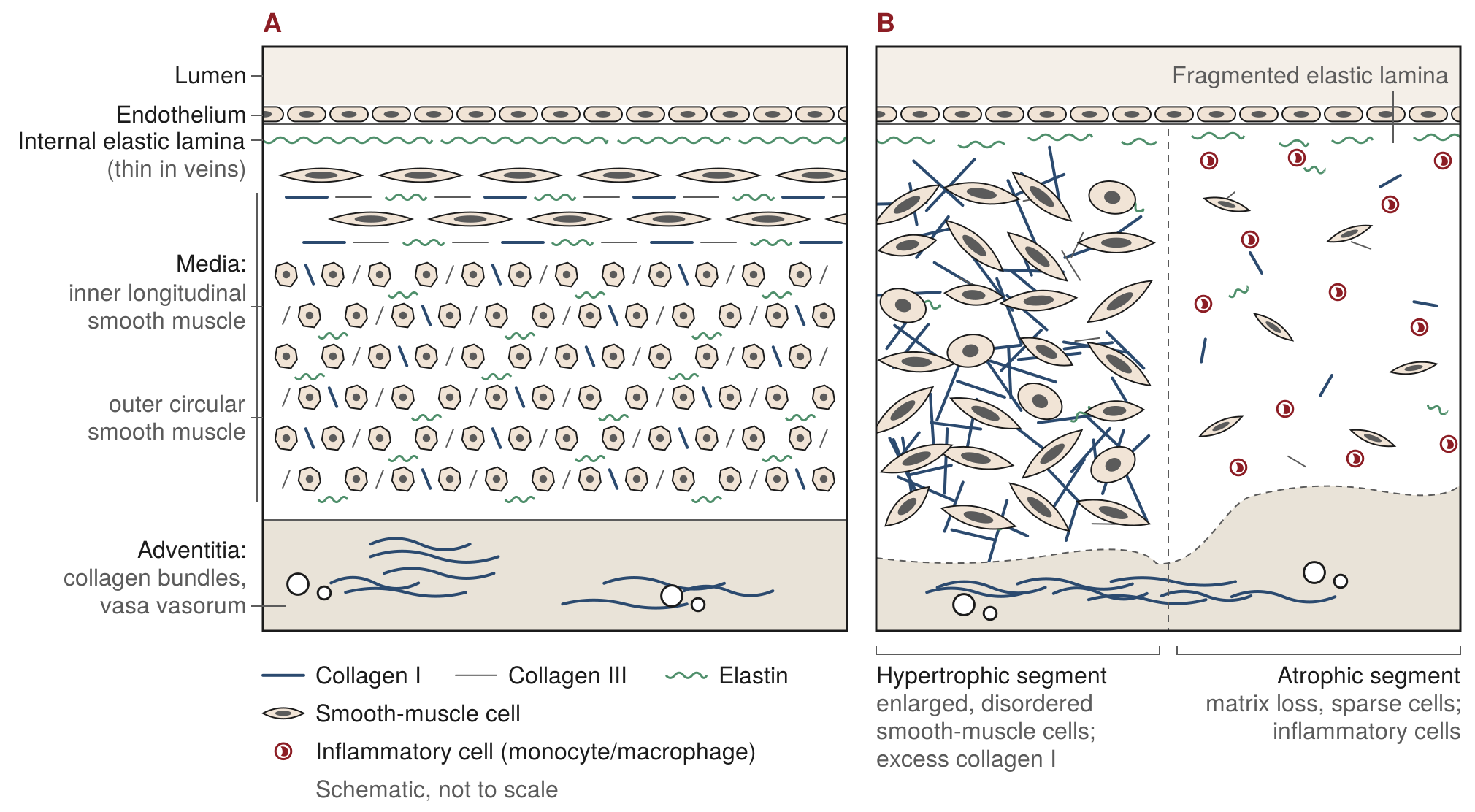
Collagen is part of the varicose vein story, but it is not the whole story. Varicose vein walls are structurally different from normal ones: the balance of collagen types is shifted, elastic fibres are fragmented and the muscle layer is disorganised. Wall changes and valve failure appear to feed each other rather than one simply causing the other.
For most people, this is a tendency built into the vein wall by many genes acting together, not a single collagen disorder. Only one rare inherited condition, vascular Ehlers–Danlos syndrome, has clear data linking a collagen gene to early varicose veins, and it changes the treatment plan. Being flexible, or having soft skin, does not.
In one sentence
The vein wall, not only the valve, is part of varicose vein disease; most people inherit that tendency through many small genetic effects, a very few have a true collagen disorder that must be recognised before treatment, and ordinary flexibility is neither a diagnosis nor a barrier to treatment.
What collagen is, and why a vein needs it
Collagen is the main structural protein of the body — the fibre that gives skin its strength, tendons their pull and bone its framework. Humans make at least 28 types, each with its own job.1 Two matter most for veins.
Type I collagen is the most abundant. Its fibres are thick and strong, like the rope in a climbing harness. Type III collagen forms finer, more pliable fibres, woven together with type I in the skin and in the walls of blood vessels, and is thought to let a tissue stretch and spring back without losing strength.1 Alongside them sits elastin, a protein that behaves like a rubber band, letting a vein widen when it fills and recoil when it empties.
A useful picture is a garden hose. The rubber is elastin: it gives. The braided mesh in the wall is collagen: it stops the hose ballooning when the pressure rises. Too little mesh, or mesh of the wrong weave, and the hose bulges; too little rubber, and it no longer springs back.
The cells that make collagen — fibroblasts in the skin and smooth muscle cells in the vein wall — plait three protein strands into a rope-like triple helix, and the enzymes that stabilise that plait cannot work without vitamin C.2 Collagen is also long-lived: half of the collagen in skin is replaced only about every 15 years.3 Whatever damage the fibres accumulate — from sun, sugar or smoke — therefore tends to stay.
Why a vein wall can weaken: the wall and the valve
For decades, varicose veins were explained in one sentence: a valve fails, blood falls backwards, the vein stretches. Valves certainly matter, and I explain how reflux (backward flow) develops in what actually causes varicose veins. But when researchers looked closely at veins removed at surgery, the wall itself turned out to be abnormal.
What the varicose wall looks like
Under the microscope, a varicose vein is not simply a stretched normal vein. It is patchy: some segments are thickened, with enlarged, disorganised muscle cells and excess matrix; others are thinned, with matrix breakdown and inflammatory cells. The elastic fibres are reduced and fragmented.4
The collagen balance shifts
The most consistent chemical finding is a change in the balance of collagen types. Smooth muscle cells grown from varicose veins make more type I collagen and less type III than cells from normal veins.5 A separate, small study that measured more than 150 matrix proteins directly in vein tissue — six varicose veins against six normal ones — found the same pattern: roughly two and a half times more type I and about a quarter of the type III.6
That is a hose with more rope and less spring, which may explain why varicose veins are both more easily stretched and, oddly, stiffer than normal veins.4 The type III deficit is not a failure of the cell’s instructions: the protein is made normally but is then broken down by enzymes in the surrounding matrix.7
A whole-body tendency?
In a separate experiment, the same French group grew skin cells from people with varicose veins and found a similar imbalance in the skin.8 That raised the possibility that the tendency is body-wide and inherited, rather than confined to a stretched vein. Two cautions. These were small laboratory studies from one group, not independently repeated. And when the group later reported the same pattern in skin from patients with venous ulcers, the skin had been sampled from the groin, close to the diseased veins — which, as they acknowledged, could partly explain the finding without any body-wide change.9 The whole-body idea is plausible, but it remains a hypothesis.
Which comes first?
A 1993 study measured collagen and elastin in varicose veins, in normal veins, and in a handful of still-competent segments from legs with varicose veins elsewhere. The competent segments had the same abnormal collagen-to-elastin ratio as the varicose ones.10 Only four such segments were studied, so this suggests, rather than proves, that wall changes come first. Others have noted varicose veins developing below valves that still work.4
The valve-first view has its own evidence: inflammatory cells crowding damaged valve leaflets, and the way reflux spreads down a vein once a valve gives way. Nor does every laboratory finding agree; some studies report more collagen in varicose veins, some less, some the same.4 My reading is that both are true. Wall and valves are part of the same disease, and once reflux begins, the rise in pressure and inflammation feeds back on both. The wall is part of the problem, not all of it.
What the genes say
If ordinary varicose veins were a mild collagen disorder, the collagen genes behind conditions such as Ehlers–Danlos syndrome would be expected to show up in large genetic studies. They do not appear among the main signals. A 2022 study of more than 135,000 people with varicose veins found 46 risk regions in the genome; some involve matrix proteins (EFEMP1, COL27A1), but the leading signals across these studies concern how the vein senses flow, how vessels develop and how valves form.11 Common varicose veins are a many-genes condition in which the wall is one of several players, as I explain in the genetics of varicose veins.
Conditions that affect collagen and connective tissue
The Ehlers–Danlos syndromes
Ehlers–Danlos syndrome, usually shortened to EDS, is not one condition but a family of them. The 2017 international classification recognises 13 types, each with its own features and, for most, its own gene.12 Three come up most often.
Classical EDS is caused by a fault in type V collagen, a minor collagen that helps set the thickness of type I fibres. The skin is noticeably stretchy and soft, scars are wide and thin like cigarette paper, and the joints are loose. It affects roughly one person in 20,000.13 In a review of 122 people with classical EDS, varicose veins were recorded in seven, about 6%, which is not clearly above the general population.14
Hypermobile EDS is the commonest type. Its hallmark is generalised joint hypermobility with pain and other symptoms; the skin is often unusually soft or velvety. Despite its name, no gene has been found for hypermobile EDS, and no blood test confirms it.15 People with symptomatic hypermobility who do not meet the full criteria are said to have a hypermobility spectrum disorder. For both, no controlled ultrasound study has ever measured how common varicose veins are, so nobody can say whether the rate is higher than in anyone else; the specialist reference on hypermobile EDS lists varicose veins among the “non-specific” findings, common in the general population.15
Vascular EDS is different, and it is the type a vein surgeon must know about. It is caused by a fault in the gene for type III collagen (COL3A1), the pliable collagen on which vessel walls and bowel wall depend. It is rare — estimates range from about one in 10,000 to one in 150,000 — and its seriousness lies in the arteries and internal organs, which can tear. The skin is thin and translucent, so that veins show through on the chest and limbs, and bruises appear in unusual places without obvious injury.16
It is the only connective tissue disorder with proper numbers on varicose veins. In a French series of 68 people with a confirmed COL3A1 fault, 41% had superficial venous insufficiency (37% with symptoms and 4% without), against an expected 17–23% in the general population. The veins appeared early — at an average age of 26, and before 20 in 44% of those affected — and most had mild disease rather than skin damage or ulcers.17 Varicose veins appearing before 30, and not related to pregnancy, are a recognised prompt to consider the diagnosis.12,18 I return to this below.
Marfan syndrome, briefly
Marfan syndrome is often grouped with the collagen disorders, but its fault lies in fibrillin-1, the scaffolding for elastic fibres. People with Marfan syndrome are typically tall and long-limbed and need monitoring of the aorta; the specialist literature does not list varicose veins among its features.19
Collagen damage you acquire
None of the following has been shown to cause varicose veins directly, but each affects the skin over the veins, which matters for how your legs look, feel and heal.
- Age. Skin collagen falls with age and skin thins. Because collagen is replaced so slowly, sugar-derived cross-links build up on the fibres year after year, making old collagen stiffer.3,20
- Sun. Ultraviolet light switches on enzymes in the skin that break collagen down, and repair is incomplete — the mechanism of premature ageing on sun-exposed skin.21
- Smoking. In smokers’ skin, the gene for a collagen-degrading enzyme is switched on more strongly than in non-smokers’, which fits their premature wrinkling.22
- Diabetes. High blood sugar accelerates the same cross-linking that age produces; the most abundant cross-link is almost twice as high in the skin of people with diabetes in their thirties and forties.20 The result is stiffer, tougher tissue — not stronger tissue.
- Cortisone-type steroids. Glucocorticoids, used over long periods, suppress collagen production in skin cells, so the skin thins and becomes fragile.23
- Vitamin C deficiency. Because the collagen-building enzymes cannot work without vitamin C, a diet lacking it for a few months leads to scurvy: bruising, bleeding gums and wounds that will not heal.2,24
Flexible joints and soft skin: what the link is, and what it is not
Generalised joint hypermobility — joints that move further than usual — is common, more so in women and in children, and it declines with age. Most people with flexible joints have no symptoms and no condition.
So does flexibility go with varicose veins? The evidence is thin. The one study that followed hypermobile and non-hypermobile people over time examined 718 young male military recruits in Iran, of whom about 29% were hypermobile. After three months of standing duty, signs of venous insufficiency had increased in the hypermobile group but not in the others.25 It is suggestive, but it involved only young men, used no ultrasound and reported no effect sizes. An American study that followed 1,025 adults for 11 years found that each 5° of extra ankle flexibility went with an 11% higher chance of venous disease progressing, which the authors tentatively put down to joint laxity.26 That is the sum of the evidence.
Just as important is what has not been shown. Patients sometimes arrive worried that because they are “bendy”, treatment will not hold. No study has found that flexible joints, stretchy skin or any other connective tissue feature predicts whether varicose veins return after treatment. The question has simply not been studied, and I would rather say so than guess in either direction.
A simple self-check of flexibility
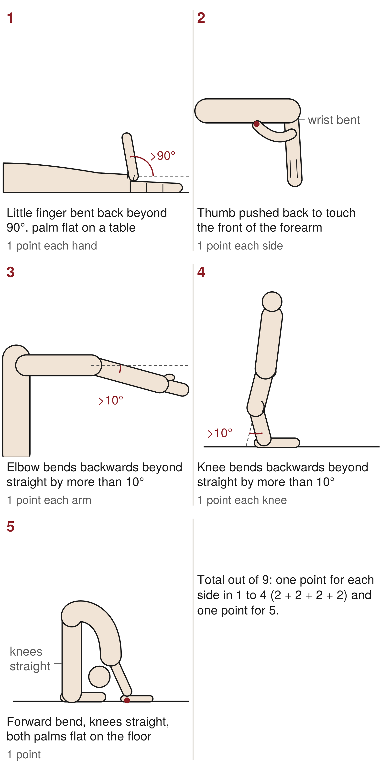
Doctors measure hypermobility with a nine-point scale called the Beighton score. It has a South African origin: it was first used in a 1973 survey of 1,081 Tswana people in Phokeng, north-west of Johannesburg.27 You can try the five manoeuvres at home.
- Little finger. Palm flat on a table, can the little finger be bent back beyond 90 degrees? One point each hand.
- Thumb. Wrist bent forward, can your thumb be pushed back to touch the front of your forearm? One point each side.
- Elbow. Arm straight, does the elbow bend backwards beyond straight by more than about 10 degrees? One point each arm.
- Knee. Standing with knees straight, do they bend backwards beyond straight by more than about 10 degrees? One point each knee.
- Forward bend. Knees straight, can you place both palms flat on the floor? One point.
The total is out of nine. The current cut-offs for generalised hypermobility are 6 or more before puberty, 5 or more from puberty to 50, and 4 or more after 50, because everyone becomes less flexible with age.15
For adults who were flexible when younger, five questions capture what the score may now miss: can you now, or could you ever, place your hands flat on the floor without bending your knees, or bend your thumb to touch your forearm? As a child, could you contort your body or do the splits? Has your shoulder or kneecap dislocated more than once? Do you consider yourself double-jointed? Two or more “yes” answers suggest hypermobility.28
This is not a diagnosis. The Beighton score was designed to survey populations, not to diagnose individuals. It is weighted towards the upper limb, leaves out many major joints, and should not be used on its own to rule hypermobility in or out.29 Hypermobile EDS requires a careful assessment of several body systems, and new international criteria for all the EDS types and for hypermobility spectrum disorders are due to be published on 1 December 2026 in the American Journal of Medical Genetics; their content is not yet public.30 If your score is high and you have symptoms — joint pain, dislocations, unusual bruising, or the skin features above — the right person to see is a rheumatologist or clinical geneticist, not a vein surgeon.
The companion observation is skin stretch, tested by gently pinching and lifting the skin on the inside of the forearm; more than about 1.5 cm is regarded as hyperextensible.13 Soft, stretchy skin is a feature of classical and hypermobile EDS, but it is also a feature of many perfectly healthy people.
From the consulting room
Our treatments are done under local anaesthetic, so a needle passing through the skin is part of almost every procedure. Some people’s skin is very soft and the needle slips through with almost no resistance; other people’s is tough and the needle needs real pressure. This is an informal impression, not something I have measured or recorded.
Soft, easily pierced skin is consistent with the “soft or velvety” skin described in hypermobile and classical EDS. But no study has measured the force needed to pierce living skin and compared it with collagen or hypermobility, so the connection is a hunch, not a finding. Tough skin is even less specific: it is more common in men; it comes with age, sun, smoking and the sugar cross-links of diabetes; and long-standing venous disease itself hardens the skin of the lower leg (lipodermatosclerosis). Tough does not mean strong — skin stiffened by glycation or scarring heals less well, not better. A needle that meets resistance tells me something about the skin, but nothing reliable about the veins beneath.
Pregnancy and hormones
Pregnancy is the most common reason for varicose veins to appear early in a woman’s life, and it has nothing to do with inherited collagen disorders. In a Finnish population study of nearly 7,000 people aged 40 to 60, the odds of varicose veins rose with each birth (odds ratios 1.2 to 2.8), while a family history was the strongest single indicator, with nearly fivefold odds.31
Several things happen at once in pregnancy: blood volume increases, pregnancy hormones are thought to relax the muscle in vein walls and make veins more distensible, pressure within the abdomen rises, and in later months the womb presses on the pelvic veins.4 No specific pregnancy hormone has been shown to weaken vein collagen. Varicose veins that appear with pregnancy are an ordinary, expected pattern, not a sign of a connective tissue disorder — which is why the warning signs below exclude them.
When to seek specialist assessment
Vascular Ehlers–Danlos syndrome is rare, and most people with early varicose veins do not have it. But please mention it to me, or to any doctor, before any vein treatment if you have:
- varicose veins that appeared before the age of 30 and were not related to pregnancy
- thin, see-through skin in which the veins of the chest, arms or legs are unusually visible
- easy bruising, particularly in odd places such as the cheeks or back, without a clear injury
- a family history of sudden death at a young age, or of a burst or torn artery in a young relative
- a personal or family history of a ruptured bowel, a ruptured womb in late pregnancy, or a collapsed lung without obvious cause
Early varicose veins are a recognised prompt for genetic testing of the type III collagen gene.16,18 If vascular EDS is suspected, a clinical geneticist is involved first and the plan for the veins is adjusted as described below.32
What this means for treatment
Everyone has a scan
Every patient at our three centres has a duplex ultrasound scan before any treatment is planned. Collagen biology can explain why a vein wall is susceptible; it cannot show which vein is leaking, where the reflux starts or how far it extends. The scan can. Treatment is directed at the source, most often with radiofrequency ablation under local anaesthetic, medical adhesive closure or ultrasound-guided foam sclerotherapy, depending on the anatomy. We treat around 750 patients a year with radiofrequency ablation across the three centres.
Ordinary flexibility: no evidence that treatment works less well
If you are hypermobile, have soft skin, or carry a diagnosis of hypermobile EDS or a hypermobility spectrum disorder, your veins are assessed and treated on the same basis as anyone else’s. There is no evidence that treatment is less effective. Whether your veins are more likely to return has simply not been studied. Bruising and slow healing of the small access wounds have been reported in EDS, but only in individual case reports.
Varicose veins can come back after any treatment. In randomised trials with at least two years of follow-up, about 22% of legs had recurrent varicose veins after endovenous ablation — the same rate as after conventional surgery. The single largest reason was the treated vein partially reopening (about a third of recurrences), followed by reflux in a neighbouring vein not treated the first time.33 Whether a connective tissue tendency adds to that, no one knows. I discuss why veins return under recurrent varicose veins.
Vascular EDS changes the plan
For the small number of people with vascular EDS, the plan is different, because the vessels are fragile. In the French series, nine patients had undergone conventional open surgery — tying off and stripping the saphenous vein in the groin — before their diagnosis was known. Two suffered serious vascular complications (a torn femoral vein in one, life-threatening bleeding in another), and the varicose veins came back in all nine. The six treated after diagnosis, once compression was not enough, with foam sclerotherapy (five) or radiofrequency ablation (one) had no vascular complications.17
The international consensus on vascular EDS therefore advises that vein stripping be avoided and that compression is the first-line treatment; endovenous ablation has been reported as successful, and foam sclerotherapy as a complementary or alternative option, when compression is not enough.32 Elective surgery of any kind is approached with caution, with the geneticist involved in the decision.16 The numbers are small, but they all point the same way.
What you can do
You cannot change the collagen you inherited, and no treatment restores the collagen balance of a vein wall. There are still sensible things to do.
- Wear compression when it is appropriate. Compression stockings reduce aching and swelling, help during pregnancy and long periods of standing, and are the first-line treatment for venous insufficiency in vascular EDS.32
- Keep walking. The calf muscle pump returns blood from your legs; regular walking, and avoiding long spells of standing still or sitting, support it whatever your collagen.
- Keep a healthy weight. Excess weight raises the pressure in the leg veins and worsens symptoms in established disease.
- Protect your skin. Sun and smoking degrade skin collagen, and ageing and diabetes stiffen it.20–22 Sun protection, not smoking and good blood sugar control will not reverse varicose veins, but they preserve the skin that has to heal after any treatment.
- Eat enough vitamin C. Your collagen-building enzymes need it,2 and a normal diet with fruit and vegetables provides plenty.
- Be sceptical of collagen supplements for veins. Swallowed collagen is broken down in the gut like any other protein, and I am aware of no study showing that collagen powders or capsules strengthen vein walls, prevent varicose veins or reduce recurrence. There is no evidence that they help veins.
- Tell your doctor the whole story. Family history, pregnancies, joint problems, unusual bruising and the warning signs above cost nothing to mention and occasionally change everything.
From the consulting room
When a patient tells me they are double-jointed, I take it as useful information rather than a verdict. Every patient has a duplex ultrasound scan before treatment, and it is the scan that shows which veins are leaking and what needs treating. Only rarely does a patient’s connective tissue change the plan; for most people, flexibility is a fact about them, not a diagnosis.
Bottom line
Collagen and the vein wall are part of varicose vein disease, and most people inherit that tendency through many small genetic effects rather than one faulty gene. Flexible joints and soft skin are common; they have not been shown to cause varicose veins, and there is no evidence that they stop treatment working. The rare exception, vascular Ehlers–Danlos syndrome, has warning signs worth knowing — and in every case the duplex scan, not the collagen, decides the treatment.
Frequently asked questions
Are varicose veins a collagen disorder?
Not in the usual sense of the term.
The wall of a varicose vein does show abnormal collagen and fragmented elastic fibres. But ordinary varicose veins are a many-genes condition in which valves, flow sensing and the wall all contribute, and the genes behind true collagen disorders are not among the main risk genes.
I am double-jointed. Does that mean I will get varicose veins?
Not necessarily.
Hypermobility is common, and most hypermobile people never develop a connective tissue disorder. Two modest studies hint at a link with venous disease, but no controlled ultrasound study has shown that hypermobile people have more varicose veins.
Will my varicose veins come back after treatment because I have hypermobile EDS?
Nobody knows.
Varicose veins can return after any treatment, in about one in five legs over a few years, most often because the treated vein partly reopens or a neighbouring vein starts to leak. Whether a connective tissue tendency adds to that risk has never been studied.
Do collagen supplements help varicose veins?
There is no evidence that they do.
Swallowed collagen is broken down in the gut like any other protein, and no study has shown that collagen powders or capsules strengthen vein walls, prevent varicose veins or reduce recurrence.
Is a high Beighton score a diagnosis of Ehlers–Danlos syndrome?
No.
The Beighton score describes flexibility; it was designed for surveying populations and leaves out many joints. A diagnosis of hypermobile EDS requires a full assessment by a rheumatologist or clinical geneticist.
Can I still have vein treatment if I have a connective tissue disorder?
Usually, yes.
People with hypermobile or classical EDS, or a hypermobility spectrum disorder, are treated in the same way as anyone else. Vascular EDS is the exception: open vein stripping should be avoided, and the approach is compression first, then foam sclerotherapy or endovenous ablation if needed, planned with the geneticist.
My skin is thin and I bruise easily. Should I be worried?
Usually not, but it is worth mentioning.
Thin skin and bruising are common with age, sun and steroid use. They matter when they occur together with varicose veins before 30 unrelated to pregnancy, see-through skin, or a family history of a burst artery, bowel or womb, or of sudden death at a young age. Then ask for assessment before any vein treatment.
References
- Ricard-Blum S. The collagen family. Cold Spring Harb Perspect Biol. 2011;3(1):a004978. doi:10.1101/cshperspect.a004978
- Gjaltema RAF, Bank RA. Molecular insights into prolyl and lysyl hydroxylation of fibrillar collagens in health and disease. Crit Rev Biochem Mol Biol. 2017;52(1):74–95. doi:10.1080/10409238.2016.1269716
- Verzijl N, DeGroot J, Thorpe SR, et al. Effect of collagen turnover on the accumulation of advanced glycation end products. J Biol Chem. 2000;275(50):39027–39031. doi:10.1074/jbc.M006700200
- Raffetto JD, Khalil RA. Mechanisms of lower extremity vein dysfunction in chronic venous disease and implications in management of varicose veins. Vessel Plus. 2021;5:36. doi:10.20517/2574-1209.2021.16
- Sansilvestri-Morel P, Rupin A, Badier-Commander C, et al. Imbalance in the synthesis of collagen type I and collagen type III in smooth muscle cells derived from human varicose veins. J Vasc Res. 2001;38(6):560–568. doi:10.1159/000051092
- Barallobre-Barreiro J, Oklu R, Lynch M, et al. Extracellular matrix remodelling in response to venous hypertension: proteomics of human varicose veins. Cardiovasc Res. 2016;110(3):419–430. doi:10.1093/cvr/cvw075
- Sansilvestri-Morel P, Rupin A, Jullien ND, et al. Decreased production of collagen type III in cultured smooth muscle cells from varicose vein patients is due to a degradation by MMPs: possible implication of MMP-3. J Vasc Res. 2005;42(5):388–398. doi:10.1159/000087314
- Sansilvestri-Morel P, Rupin A, Jaisson S, et al. Synthesis of collagen is dysregulated in cultured fibroblasts derived from skin of subjects with varicose veins as it is in venous smooth muscle cells. Circulation. 2002;106(4):479–483. doi:10.1161/01.CIR.0000022846.22923.46
- Labropoulos N, Sansilvestri-Morel P, Kerihuel JC, et al. III – Pathophysiology [congress report: guest lecture, satellite workshop “Tissular repair in phlebology” and venous pathophysiology session]. Phlebolymphology. 2010;17(1), No. 66. https://phlebolymphology.org/?p=3310
- Gandhi RH, Irizarry E, Nackman GB, et al. Analysis of the connective tissue matrix and proteolytic activity of primary varicose veins. J Vasc Surg. 1993;18(5):814–820.
- Ahmed WUR, Kleeman S, Ng M, et al. Genome-wide association analysis and replication in 810,625 individuals with varicose veins. Nat Commun. 2022;13:3065. doi:10.1038/s41467-022-30765-y
- Malfait F, Francomano C, Byers P, et al. The 2017 international classification of the Ehlers-Danlos syndromes. Am J Med Genet C Semin Med Genet. 2017;175(1):8–26. doi:10.1002/ajmg.c.31552
- Malfait F, Symoens S, Syx D. Classic Ehlers-Danlos syndrome. In: Adam MP, et al., editors. GeneReviews®. Seattle (WA): University of Washington; updated 1 February 2024.
- D'hondt S, Van Damme T, Malfait F. Vascular phenotypes in nonvascular subtypes of the Ehlers-Danlos syndrome: a systematic review. Genet Med. 2018;20(6):562–573. doi:10.1038/gim.2017.138
- Hakim A. Hypermobile Ehlers-Danlos syndrome. In: Adam MP, et al., editors. GeneReviews®. Seattle (WA): University of Washington; updated 22 February 2024.
- Byers PH. Vascular Ehlers-Danlos syndrome. In: Adam MP, et al., editors. GeneReviews®. Seattle (WA): University of Washington; updated 10 April 2025.
- Frank M, Says J, Denarié N, Messas E. Histoire naturelle de l’insuffisance veineuse superficielle chez les patients atteints de syndrome d’Ehlers-Danlos vasculaire [Natural history of superficial venous insufficiency in patients with vascular Ehlers-Danlos syndrome]. Phlébologie. 2015;68(3):34–40.
- Bowen JM, Hernandez M, Johnson DS, et al. Diagnosis and management of vascular Ehlers-Danlos syndrome: experience of the UK national diagnostic service, Sheffield. Eur J Hum Genet. 2023;31(7):749–760. doi:10.1038/s41431-023-01343-7
- Dietz H. FBN1-related Marfan syndrome. In: Adam MP, et al., editors. GeneReviews®. Seattle (WA): University of Washington; updated 17 February 2022.
- Monnier VM, Sun W, Sell DR, Fan X, Nemet I, Genuth S. Glucosepane: a poorly understood advanced glycation end product of growing importance for diabetes and its complications. Clin Chem Lab Med. 2014;52(1):21–32. doi:10.1515/cclm-2013-0174
- Fisher GJ, Wang ZQ, Datta SC, Varani J, Kang S, Voorhees JJ. Pathophysiology of premature skin aging induced by ultraviolet light. N Engl J Med. 1997;337(20):1419–1428. doi:10.1056/NEJM199711133372003
- Lahmann C, Bergemann J, Harrison G, Young AR. Matrix metalloproteinase-1 and skin ageing in smokers. Lancet. 2001;357(9260):935–936. doi:10.1016/S0140-6736(00)04220-3
- Schoepe S, Schäcke H, Asadullah K. Test systems for the determination of glucocorticoid receptor ligand induced skin atrophy. Dermatoendocrinol. 2011;3(3):175–179. doi:10.4161/derm.3.3.15065
- Gandhi M, Elfeky O, Ertugrul H, Chela HK, Daglilar E. Scurvy: rediscovering a forgotten disease. Diseases. 2023;11(2):78. doi:10.3390/diseases11020078
- Azma K, Mottaghi P, Hosseini A, Salek S, Bina R. Venous insufficiency after prolonged standing: is joint hypermobility an important risk factor? Adv Biomed Res. 2015;4:98. doi:10.4103/2277-9175.156666
- Kim TI, Forbang NI, Criqui MH, Allison MA. Association of foot and ankle characteristics with progression of venous disease. Vasc Med. 2015;20(2):105–111. doi:10.1177/1358863X14568443
- Beighton P, Solomon L, Soskolne CL. Articular mobility in an African population. Ann Rheum Dis. 1973;32(5):413–418. doi:10.1136/ard.32.5.413
- Hakim AJ, Grahame R. A simple questionnaire to detect hypermobility: an adjunct to the assessment of patients with diffuse musculoskeletal pain. Int J Clin Pract. 2003;57(3):163–166.
- Malek S, Reinhold EJ, Pearce GS. The Beighton Score as a measure of generalised joint hypermobility. Rheumatol Int. 2021;41(10):1707–1716. doi:10.1007/s00296-021-04832-4
- The Ehlers-Danlos Society. A major update is coming to EDS and HSD diagnosis: new global diagnostic criteria. 11 March 2026 (updated 12 June 2026). https://www.ehlers-danlos.com/new-global-diagnostic-criteria-2026/ (accessed 7 November 2026).
- Laurikka JO, Sisto T, Tarkka MR, Auvinen O, Hakama M. Risk indicators for varicose veins in forty- to sixty-year-olds in the Tampere varicose vein study. World J Surg. 2002;26(6):648–651. doi:10.1007/s00268-001-0283-1
- Byers PH, Belmont J, Black J, et al. Diagnosis, natural history, and management in vascular Ehlers-Danlos syndrome. Am J Med Genet C Semin Med Genet. 2017;175(1):40–47. doi:10.1002/ajmg.c.31553
- O’Donnell TF, Balk EM, Dermody M, Tangney E, Iafrati MD. Recurrence of varicose veins after endovenous ablation of the great saphenous vein in randomized trials. J Vasc Surg Venous Lymphat Disord. 2016;4(1):97–105. doi:10.1016/j.jvsv.2014.11.004