Lipodermatosclerosis is an inflammatory and fibrosing complication of chronic venous hypertension, classed as CEAP C4b disease. Its acute phase is regularly mistaken for cellulitis. This review covers how common it is, what happens in the tissue, how to recognise and investigate it, and what current European and North American guidelines recommend.
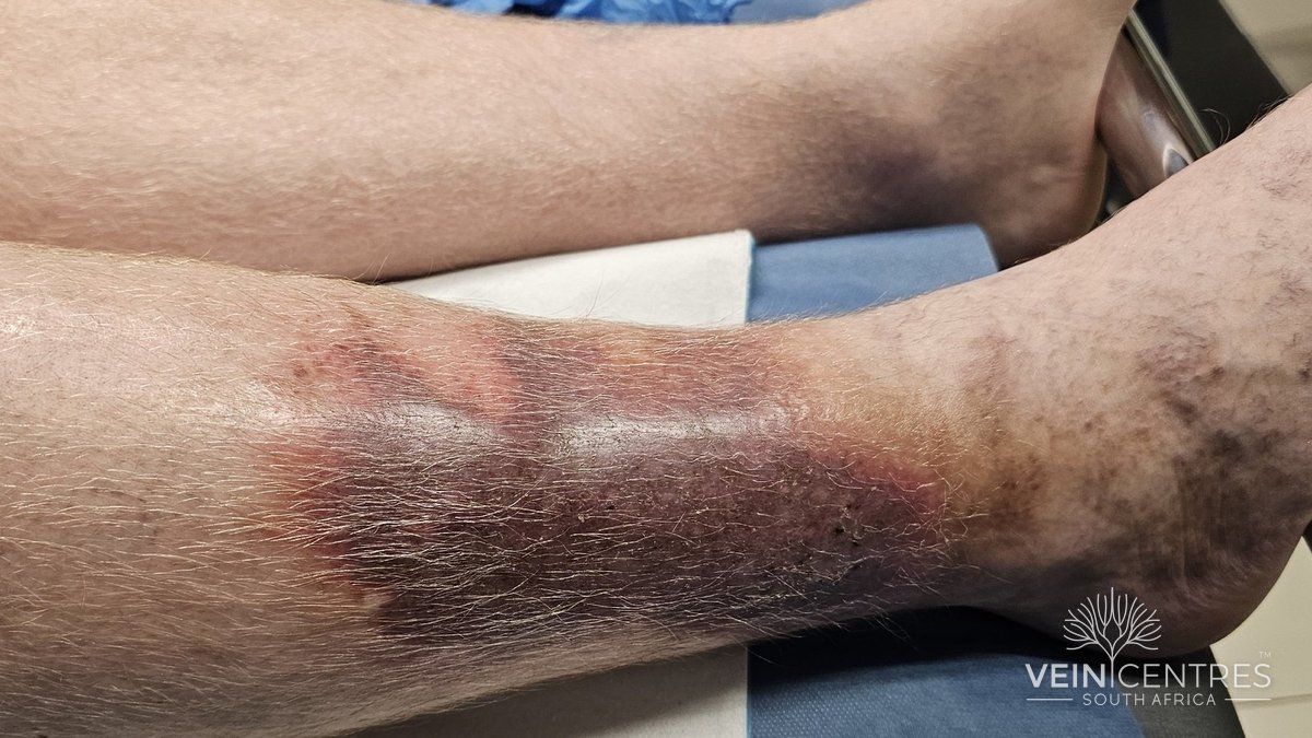
The patient on her third course of antibiotics
A composite case, typical of referrals to a vein practice
She is 58, a teacher who stands most of the day, with a body-mass index of 33. For six weeks, the skin above her left ankle has been red, warm and exquisitely tender. At times, even the weight of the bedsheet hurts.
She has already had flucloxacillin, then amoxicillin–clavulanate, followed by intravenous cefazolin in hospital. She has never been febrile and has remained systemically well. Her inflammatory markers have never convincingly suggested infection, and the redness has not behaved like cellulitis.
When she finally sees a vein surgeon, the other leg provides the clue: brown haemosiderin pigmentation around the ankle, firm induration of the lower calf and early narrowing of the leg immediately above the malleolus. A venous duplex scan shows extensive great saphenous vein reflux.
The diagnosis is not recurrent cellulitis. It is lipodermatosclerosis (LDS), an inflammatory and fibrosing manifestation of chronic venous disease. By the time the characteristic skin changes have appeared, the underlying venous disease is already advanced.
What is lipodermatosclerosis?
Lipodermatosclerosis is inflammation and fibrosis of the skin and subcutaneous fat of the lower leg, usually associated with longstanding venous hypertension. It has also been called sclerosing panniculitis and hypodermitis sclerodermiformis.
In the 2020 CEAP classification, venous skin changes are divided into three subclasses:5
- C4a — pigmentation or eczema
- C4b — lipodermatosclerosis or atrophie blanche
- C4c — corona phlebectatica
LDS is therefore C4b disease. It is not a cosmetic consequence of varicose veins. It indicates established tissue damage and a leg at increased risk of venous ulceration.
The literature describes three clinical stages, which often overlap.13
| Stage | Duration | Typical picture |
|---|---|---|
| Acute | <1 month | Painful, red, warm, poorly defined induration |
| Subacute | 1–12 months | Less tender, increasingly pigmented and indurated |
| Chronic | >1 year | Woody fibrosis, pigmentation, narrowed lower calf |
How common is it?
The incidence of LDS itself is unknown. Population studies report the broader CEAP C4 category, which also includes pigmentation, eczema and corona phlebectatica, so C4 figures are a proxy rather than a measure of LDS. Even so, advanced venous skin changes are not rare:
- The Bonn Vein Study examined 3072 adults aged 18 to 79 years and found C4 skin changes in 2.9% (3.1% of men and 2.7% of women).7
- A global systematic review of 32 studies from six continents, covering more than 300,000 adults, estimated the pooled C4 prevalence at about 4%.6
Almost all of these data come from European and North American populations. There are virtually no population data from sub-Saharan Africa, including South Africa.
Who develops LDS?
The best clinical description remains a Mayo Clinic series of 97 patients.8
| Feature | Finding |
|---|---|
| Women | 87% |
| Mean age (range) | 62 yr (25–88) |
| Mean body-mass index | 34.3 |
| Bilateral disease | 45% |
| Oedema | 71% |
| Hyperpigmentation | 59% |
| Ulceration | 13% |
Of the 72 patients who had vascular studies, 49 had abnormal results. Deep venous incompetence was the commonest finding (67% of abnormal studies), followed by calf-muscle pump dysfunction (39%).8 LDS is therefore not only a superficial-vein problem.
Obesity, age, prolonged standing, previous deep-vein thrombosis, poor calf-muscle pump function and superficial or deep venous incompetence all raise venous pressure and therefore risk.
What is happening underneath the skin?
The simplest summary is venous hypertension → microvascular injury and inflammation → fibrosis. The biology is more complicated, and the mechanisms below are best seen as overlapping models rather than a settled sequence.13
Venous hypertension. Normally the calf-muscle pump and competent valves lower venous pressure in the leg during walking. With superficial or deep reflux, or outflow obstruction, ambulatory venous pressure stays high and is transmitted into the skin microcirculation.
Microvascular leakage. Dermal capillaries become distended and permeable. Red cells and plasma proteins escape into the tissue, and the breakdown of red cells deposits haemosiderin, producing the brown pigmentation of the lower leg.
Endothelial activation and inflammation. Leucocyte trapping and activation, endothelial dysfunction and the release of inflammatory mediators create an inflammatory microenvironment in the skin and fat.13 LDS is therefore more than simple back-pressure.
Fibrin deposition. Pericapillary fibrin was found in all LDS skin examined in the classic studies.11 The original “fibrin cuff” hypothesis — that fibrin blocks oxygen diffusion — is no longer thought sufficient on its own, but abnormal fibrin turnover and impaired fibrinolysis remain part of the picture.
Fibrosis. Activated leucocytes release transforming growth factor β1, which drives fibroblasts to lay down collagen; active TGF-β1 levels are markedly raised in C4 skin.12 The subcutaneous fat becomes inflamed (panniculitis) and is progressively replaced by fibrous tissue.
Lymphatic failure. In advanced disease the lymphatics are overloaded and then damaged, adding protein-rich oedema and further inflammation (phlebolymphoedema).13
The end result is the classic inverted champagne-bottle leg: a broad upper calf narrowing sharply into a hard, contracted lower leg. At this stage the skin is poorly compliant, vulnerable to minor trauma and increasingly prone to ulceration.
Clinical presentation
Acute LDS
Acute LDS can look strikingly inflammatory. The typical site is the medial lower leg above the ankle, and patients may have intense pain and marked tenderness, erythema and warmth, swelling, and poorly defined induration. The pain can be severe enough that compression stockings, or even light touch, are hard to tolerate at first. This is the phase most often mistaken for cellulitis.
Subacute and chronic LDS
With time, the inflamed tissue becomes increasingly fibrotic. Typical findings include:
- brown or red-brown pigmentation
- firm, woody, non-pitting induration and bound-down skin
- narrowing of the lower calf
- atrophie blanche in some patients
- venous eczema and oedema
- healed or active venous ulceration
The acute phase may precede chronic LDS, or flare repeatedly on top of established disease (“acute-on-chronic” LDS).13
LDS in darker skin
Most textbook images of LDS show pale skin, in which inflammation looks red. In darker skin, erythema is much harder to see. Active inflammation may appear violaceous, dark brown or grey, and pigmentation can become very dark. Warmth, tenderness and induration on palpation therefore matter more than colour, and the diagnosis should never rest on looking for redness.
The great mimic: LDS or cellulitis?
Cellulitis is common, but it is frequently overdiagnosed. Two 2023 meta-analyses put the size of the problem in numbers:
- In primary and urgent care, a mean of 41% of patients initially diagnosed with uncomplicated cellulitis received an alternative diagnosis on reassessment within 14 days, although individual studies ranged from 19% to 83%.16
- Among 858 inpatients, 39% were reclassified by a dermatologist or infectious diseases specialist; stasis dermatitis was the commonest alternative diagnosis.17
LDS belongs to this group of venous and inflammatory mimics.
| Favours LDS | Favours cellulitis |
|---|---|
| Longstanding venous disease, varicose veins, chronic ankle oedema | Rapid progression over hours to a few days |
| Haemosiderin pigmentation, venous eczema | Obvious portal of entry (wound, interdigital tinea) |
| Firm subcutaneous induration; inverted champagne-bottle leg | Progressively spreading erythema; lymphangitis |
| Recurrent episodes in the same area; bilateral changes | Fever, rigors, systemic illness |
| Patient systemically well | Substantially raised inflammatory markers |
| No improvement with appropriate antibiotics | Convincing response to antibiotics |
There is considerable overlap. A normal white-cell count does not exclude cellulitis, and mild elevation of C-reactive protein occurs with non-infectious inflammation, so the diagnosis cannot rest on one laboratory result.
One particularly useful clue: true simultaneous bilateral lower-leg cellulitis is uncommon. When both legs are red, warm and swollen, consider venous disease, stasis dermatitis, LDS, lymphoedema and drug reactions before labelling it bilateral cellulitis. Bilateral infection does occur, particularly in some immunocompromised patients, so bilaterality is a prompt to reconsider rather than an absolute exclusion.18
The wider differential
Beyond cellulitis, consider other panniculitides (erythema nodosum, usually on the anterior shins; erythema induratum, on the calves); fibrosing conditions such as morphea and scleroderma; superficial thrombophlebitis, deep-vein thrombosis, lymphoedema and lipoedema; contact dermatitis, often to topical treatments or bandages; and, less commonly, vasculitis, pretibial myxoedema, calciphylaxis and malignancy.
The oncology patient: another trap
Several anticancer agents can cause inflammatory reactions that resemble cellulitis, and gemcitabine is the most important. In a 2023 oncodermatology series of patients diagnosed with pseudocellulitis, acute LDS was the commonest diagnosis (74.2%), most cases were gemcitabine-associated, and about a third of patients had cancer treatment held because of presumed infection.19 A 2026 report again described gemcitabine-induced pseudocellulitis, with onset typically 2 to 7 days after each infusion.20
The practical lesson: when lower-leg inflammation recurs in step with chemotherapy cycles, without systemic signs of infection, consider pseudocellulitis before prescribing repeated antibiotics or interrupting cancer treatment.
How is lipodermatosclerosis diagnosed?
LDS is primarily a clinical diagnosis. Investigation is not mainly to prove that the skin is fibrotic. It is to find out why it became fibrotic, and whether the cause can be treated.
Venous duplex ultrasound
This is the key investigation. A complete study should assess the deep venous system, including evidence of previous thrombosis or obstruction; the great and small saphenous systems and relevant tributaries; the distribution of reflux; and perforating veins close to the area of skin change.4,13 If symptoms or duplex findings suggest proximal obstruction, add abdominal duplex and, where needed, CT or MR venography or intravascular ultrasound. Consider lymphoscintigraphy if phlebolymphoedema is suspected.4
Arterial assessment before compression
Moderate or high compression should never be applied blindly. Check pedal pulses and the vascular history, and assess the arterial supply first. ESVS advises that sustained compression requires an ankle pressure above 60 mm Hg and an ankle–brachial index above 0.6.1 Many wound-care protocols use more conservative thresholds, so local protocols and the clinical context should guide treatment.
| Source | Threshold | Compression |
|---|---|---|
| ESVS 20221 | ABI >0.6 and ankle pressure >60 mm Hg | Required before sustained compression; below this, compression is contraindicated |
| Common wound-care protocols | ABPI >0.8 | Full compression |
| Common wound-care protocols | ABPI about 0.5–0.8 | Modified (reduced) compression, ideally with specialist input |
| All settings | ABPI >1.3 | Vessels may be incompressible (diabetes, renal disease); use toe pressures or waveform analysis |
Blood tests
There is no blood test for LDS. White-cell count and C-reactive protein are useful mainly when infection is still in the differential, and should support the clinical assessment, not decide it.
Skin ultrasound
High-frequency skin ultrasound is a useful adjunct in difficult cases. In one small study, affected skin had a mean dermal thickness of 2.63 mm, as compared with 1.45 mm in healthy skin, with increased echogenicity and loss of the normal dermo-hypodermal interface.21 Standard vascular probes also show two patterns: a scleroedematous pattern in acute LDS and a fibrosclerotic pattern in chronic LDS.22 For now this is a specialised adjunct rather than a routine test.
Be cautious about biopsy
Histology can confirm LDS, but biopsy is rarely needed when the clinical picture and venous assessment are typical. Fibrotic venous skin heals poorly, and a biopsy wound can itself become a chronic ulcer. Reserve biopsy for atypical cases in which vasculitis, morphea, another panniculitis or malignancy must be excluded.13
Does the size of the LDS area matter?
Probably. In the Mayo series, 13% of patients already had ulceration.8 In a randomised compression trial involving patients with healed venous ulcers, those who had re-ulceration within two years started with a much larger area of LDS — about 293 cm², as compared with 50 cm² in those whose ulcers stayed healed.23 This cannot predict an individual patient’s risk precisely, but extensive LDS should be taken seriously. Recording the affected area with standardised photography at baseline helps follow-up and patient education.
What do current guidelines recommend?
ESVS 2022
The European Society for Vascular Surgery guideline is the only major guideline with a recommendation written specifically for LDS:1
ESVS recommendation · C4b disease
For patients with chronic venous disease and lipodermatosclerosis and/or atrophie blanche (CEAP C4b), below-knee elastic compression stockings exerting 20–40 mm Hg at the ankle are recommended to reduce skin induration.
Class I · Level B
More importantly, for patients with C4–C6 skin changes and superficial venous incompetence, interventional treatment of the incompetent veins is recommended, and the guideline regards it as an appropriate use of health care resources.1 The distinction matters. Compression treats the consequences of venous hypertension; when treatable superficial reflux is present, intervention addresses a source of it.
Other ESVS points relevant to LDS:1
- Endovenous thermal ablation is the first choice for great and small saphenous incompetence, in preference to stripping or foam (Class I, Level A).
- In C4b skin, adjacent phlebectomy may be complicated by delayed wound healing; ultrasound-guided foam sclerotherapy is therefore a useful alternative for tributaries.
- With advanced skin changes (C4b–C6), treatment of isolated or residual significant incompetent perforating veins may be considered (Class IIb, Level C). The evidence is weak and management should be individualised.
- Venoactive drugs should be considered for symptoms and oedema in patients not having, or awaiting, intervention (Class IIa, Level A).
- In active venous ulcers, early endovenous ablation is recommended to speed healing (Class I, Level B).
UIP/EVF/IUA guidelines (2018 and 2020)
The International Union of Phlebology and European Venous Forum guidelines set out the investigation of C4b disease described above. They are also the main source for the use of micronised purified flavonoid fraction (MPFF) in venous skin changes.4
SVS/AVF/AVLS (2022–2023)
The North American guidelines favour superficial venous intervention over long-term compression alone for symptomatic patients with axial superficial reflux.2,3 The revised Venous Clinical Severity Score records induration, explicitly including LDS, by its extent up the calf.
NICE Clinical Knowledge Summaries (2022)
NICE advises that potent topical glucocorticoids may be needed to settle inflammation enough for compression to be tolerated, that compression be built up gradually, and that referral for venous surgery be considered when superficial reflux is present.29
The shared principle: once significant venous skin changes have developed, prescribing stockings indefinitely without investigating the underlying venous haemodynamics is no longer adequate management.
Managing LDS in practice
Management has two objectives: control the inflammatory and oedematous consequences of venous hypertension, and correct treatable venous hypertension where possible.
1. Identify and treat the underlying venous disease
Every patient with convincing LDS deserves a proper venous assessment. Where clinically important superficial reflux is found and the patient is suitable, discuss treatment. Contemporary endovenous techniques — radiofrequency or endovenous laser ablation, ultrasound-guided foam sclerotherapy, cyanoacrylate closure and mechanochemical ablation — allow the abnormal vein to be treated without incisions through diseased lower-leg skin.1,13
Non-thermal, non-tumescent techniques can be useful when avoiding tumescent anaesthesia or thermal injury is desirable. The choice should depend on anatomy, coexisting conditions, patient preference, local expertise and the relative advantages of each technique.
Set realistic expectations. Established pigmentation and longstanding fibrosis may not resolve fully after successful treatment. The aims are to reduce venous hypertension, improve symptoms and prevent further damage and ulceration.
2. Compression remains fundamental
For chronic C4b disease, ESVS recommends 20–40 mm Hg below-knee stockings to reduce induration.1 A randomised trial showed a reduction in LDS area at 6 and 12 months in patients wearing stockings.23
Acute painful LDS is more difficult. A stocking may be intolerable when the leg is exquisitely tender. Multilayer bandaging or adjustable compression wraps can be introduced progressively, and wraps fit the irregular, narrowed lower leg better than stockings. In the 2023 series reported by Suehiro and colleagues, involving 30 patients with acute or subacute LDS, bandaging at more than 40 mm Hg relieved symptoms within about five weeks (range, 2 to 11) with only occasional analgesia. After compression stopped, 18 of 32 legs (56%) progressed to chronic LDS. The authors concluded that in these legs subcutaneous contraction is already present during the acute phase, so long-term compression is unlikely to change the prognosis.24
Compression controls venous hypertension but does not eliminate reflux. That is why the venous anatomy still needs investigating.
3. Treat venous eczema and skin inflammation
Use regular emollients and avoid irritants; use topical glucocorticoids when venous eczema is significant;29 and prescribe antibiotics only when infection is genuinely present. Settling the inflammation also makes compression easier to tolerate.
4. Drug treatment: weaker evidence than often suggested
No drug is established as first-line treatment for LDS. The evidence, from strongest to weakest, is summarised in Table 5. This article does not recommend doses for any of these agents; specialist advice should guide their use. For routine LDS, the strongest evidence remains compression plus appropriate treatment of the underlying venous disease.
| Treatment | Best evidence | Comment |
|---|---|---|
| MPFF and other venoactive drugs | Randomised trials in chronic venous disease generally, not LDS specifically4 | Reduce symptoms and oedema; useful adjunct; do not replace treatment of reflux |
| Stanozolol | Older controlled trials with compression25,26 | Hepatic, lipid, fluid-retention and androgenic effects; unavailable in many countries; not routine |
| Pentoxifylline | Randomised trials in venous ulcers; LDS data limited to small series14,15 | Effect in LDS uncertain; not established first-line therapy |
| Oxandrolone | Case reports, including a 2024 refractory ulcerated case27 | Exceptional refractory disease only |
| Hydroxychloroquine, intralesional triamcinolone, JAK inhibitors | Isolated case reports | Experimental |
5. Lifestyle
Walking, regular calf-muscle exercise, avoiding prolonged immobility, weight reduction where appropriate, leg elevation and meticulous skin care all reduce venous load. They support, but do not replace, correction of significant venous pathology.
When to refer to a vein specialist
- Suspected LDS with no previous venous duplex assessment
- C4b skin changes with varicose veins
- Recurrent episodes labelled “cellulitis” without convincing infection
- A healed or active venous ulcer
- Significant unilateral venous skin changes
- Progressive induration despite compression
- Suspected deep or proximal venous obstruction
- ABPI below 0.8, or uncertainty about arterial supply before compression
- Uncertainty about whether a red leg is infection or chronic venous disease
What has changed in the recent literature?
Venous abnormalities are common, but not universal
A 2026 Mayo Clinic study reviewed 430 patients with biopsy-confirmed LDS. Venous ultrasonography was abnormal in 327 (76%). Of the 33 patients with normal scans who underwent further haemodynamic testing, 12 had abnormal calf-muscle pump function, 7 had mild valvular incompetence and 2 had pulsatile venous flow from raised right atrial pressure. A few patients had genuinely normal haemodynamics, so other factors must also contribute.9
Two lessons follow. Venous dysfunction is present in most patients with LDS, including those with acute disease.9,10 But if the routine scan is normal, the right response is not to dismiss the diagnosis: reconsider the haemodynamics, including calf-pump function and deep venous disease, and look for other contributors such as obesity and lymphoedema.
Chemotherapy-associated pseudocellulitis
Oncology literature since 2023 has highlighted acute LDS-like reactions, particularly with gemcitabine.19,20 Recognising the pattern avoids repeated antibiotics, unnecessary admissions and inappropriate interruption of cancer treatment.
Interest in anti-inflammatory and antifibrotic treatment
Case reports of oxandrolone, pentoxifylline and immune-modulating drugs in refractory LDS show growing interest in the inflammatory and fibrotic pathways.27 These treatments remain investigational and should not distract from the underlying haemodynamic problem.
Why earlier treatment of venous hypertension makes sense
The strongest trial evidence for early venous intervention comes not from LDS but from the next stage: venous ulceration. In the EVRA trial, 450 patients with venous ulcers and superficial reflux were randomly assigned to early endovenous ablation or deferred intervention.28 The median time to ulcer healing fell from 82 to 56 days, and at 24 weeks 85.6% of ulcers had healed with early treatment, as compared with 76.3% with deferred treatment.
EVRA did not study LDS, so it cannot be claimed that early ablation in C4b disease has been proven to prevent ulcers. It does provide strong evidence for treating superficial venous hypertension once tissue damage is established — a principle reflected in the ESVS recommendation to intervene in C4–C6 disease.1
Areas of uncertainty
Honest management of LDS means being clear about what is not known.
- Does treating reflux in C4b prevent ulcers? No randomised trial has tested this; the case rests on EVRA, observational data and pathophysiology.
- Can acute LDS be stopped from becoming chronic? In the Suehiro series, compression relieved acute symptoms in every leg, yet 56% still progressed to chronic LDS.24
- How should acute LDS be distinguished from cellulitis? There are no validated diagnostic criteria, and no laboratory test reliably separates the two.
- Which drug, if any, works? Pharmacological evidence for LDS itself is limited to small or old studies and case reports.
- What drives LDS when venous studies are normal? About a quarter of biopsy-proven cases had normal routine scans.9
- When do perforating veins matter? Their contribution in C4b disease, and the benefit of treating them, remain uncertain.1
- How common is LDS? True incidence is unknown, and there are almost no data from African populations.
Key clinical points
- A red, painful lower leg is not automatically cellulitis. Around 4 in 10 initial cellulitis diagnoses are revised on review.16
- Bilateral lower-leg erythema should prompt reconsideration of the diagnosis, though it does not absolutely exclude infection.
- LDS is CEAP C4b disease — advanced chronic venous disease, not a cosmetic skin problem.
- The diagnosis is usually clinical. Duplex ultrasound identifies the underlying venous pathology, including deep venous disease.
- Check the arterial supply before compression, and avoid biopsy in severely diseased venous skin unless the diagnosis is genuinely in doubt.
- Compression is evidence-based. ESVS recommends 20–40 mm Hg below-knee stockings for C4b disease (Class I, Level B); in the acute phase, bandaging or adjustable wraps are often better tolerated.
- When C4–C6 skin changes coexist with treatable superficial venous incompetence, guidelines recommend intervention.
- Drug treatment for LDS has much weaker evidence than compression and venous intervention.
- Gemcitabine and other anticancer drugs can cause LDS-like pseudocellulitis.
- Repeated “cellulitis” that does not behave like infection deserves a venous assessment, not another automatic course of antibiotics.
A bedside rule
Red leg + chronic venous skin changes + no systemic illness + poor response to antibiotics = reconsider cellulitis and assess the venous circulation.
Disclosure. The author performs endovenous and other venous procedures in private practice. This article is for medical education and does not replace individual clinical assessment.
Clinical photographs. Vein Centres South Africa. Published with the patients’ written consent; identifying features have been removed.
How to cite
Blignaut J. Lipodermatosclerosis: The Red Leg That Isn't Cellulitis. Clinical review. Vein Centres South Africa; October 2026. Available from: https://www.veinsurgery.co.za/for-doctors/lipodermatosclerosis-clinical-review/
References
- De Maeseneer MG, Kakkos SK, Aherne T, et al. European Society for Vascular Surgery (ESVS) 2022 clinical practice guidelines on the management of chronic venous disease of the lower limbs. Eur J Vasc Endovasc Surg 2022;63:184-267.
- Gloviczki P, Lawrence PF, Wasan SM, et al. The 2022 Society for Vascular Surgery, American Venous Forum, and American Vein and Lymphatic Society clinical practice guidelines for the management of varicose veins of the lower extremities. Part I. J Vasc Surg Venous Lymphat Disord 2023;11:231-261.
- Gloviczki P, Lawrence PF, Wasan SM, et al. The 2023 Society for Vascular Surgery, American Venous Forum, and American Vein and Lymphatic Society clinical practice guidelines for the management of varicose veins of the lower extremities. Part II. J Vasc Surg Venous Lymphat Disord 2024;12:101670.
- Nicolaides A, Kakkos S, Baekgaard N, et al. Management of chronic venous disorders of the lower limbs: guidelines according to scientific evidence. Int Angiol 2018;37:181-254; Part II, Int Angiol 2020;39:175-240.
- Lurie F, Passman M, Meisner M, et al. The 2020 update of the CEAP classification system and reporting standards. J Vasc Surg Venous Lymphat Disord 2020;8:342-352.
- Salim S, Machin M, Patterson BO, Onida S, Davies AH. Global epidemiology of chronic venous disease: a systematic review with pooled prevalence analysis. Ann Surg 2021;274:971-976.
- Rabe E, Pannier-Fischer F, Bromen K, et al. Bonn Vein Study: epidemiological investigation of chronic venous diseases. Phlebologie 2003;32:1-14.
- Bruce AJ, Bennett DD, Lohse CM, Rooke TW, Davis MDP. Lipodermatosclerosis: review of cases evaluated at Mayo Clinic. J Am Acad Dermatol 2002;46:187-192.
- Garimella V, Mukosera GT, Liedl DA, et al. Venous abnormalities in biopsy-confirmed lipodermatosclerosis. Adv Skin Wound Care 2026;39:141-144.
- Greenberg AS, Hasan A, Montalvo BM, Falabella A, Falanga V. Acute lipodermatosclerosis is associated with venous insufficiency. J Am Acad Dermatol 1996;35:566-568.
- Burnand KG, Whimster I, Naidoo A, Browse NL. Pericapillary fibrin in the ulcer-bearing skin of the leg: the cause of lipodermatosclerosis and venous ulceration. Br Med J (Clin Res Ed) 1982;285:1071-1072.
- Pappas PJ, You R, Rameshwar P, et al. Dermal tissue fibrosis in patients with chronic venous insufficiency is associated with increased transforming growth factor-β1 gene expression and protein production. J Vasc Surg 1999;30:1129-1145.
- Reina-Gutierrez L, Sanjuanbenito-Reina A. Management of hypodermitis or lipodermatosclerosis: an up-to-date review. Phlebolymphology 2023;30:6-25.
- Alsararatee HH. Lipodermatosclerosis: from pathophysiology to treatment. Br J Nurs 2025;34:134-139.
- Klejtman T, et al. Specific management of lipodermatosclerosis (sclerotic hypodermitis) in acute and chronic phase. J Med Vasc 2022.
- Nightingale R, Yadav K, Hamill L, et al. Misdiagnosis of uncomplicated cellulitis: a systematic review and meta-analysis. J Gen Intern Med 2023;38:2396-2404.
- Cutler TS, Jannat-Khah DP, Kam B, Mages KC, Evans AT. Prevalence of misdiagnosis of cellulitis: a systematic review and meta-analysis. J Hosp Med 2023;18:254-261.
- Chuang YC, et al. Bilateral lower limbs cellulitis: a narrative review of an overlooked clinical dilemma. Int J Gen Med 2022.
- Pseudocellulitis in oncology patients: a single institutional retrospective analysis investigating the clinical presentation, therapeutic response, and implications for cancer therapy. JAAD Int 2023;12:186-188.
- Rusz A, Jabeen U, Zada A, Sae-Tia S. Gemcitabine-induced pseudocellulitis in metastatic pancreatic cancer. J Brown Hosp Med 2026;5:164082.
- Woźniak W, Danowska A, Mlosek RK. The use of high-frequency skin ultrasound in the diagnosis of lipodermatosclerosis. J Ultrason 2020;20:e284-e290.
- Caggiati A. Ultrasonography of skin changes in legs with chronic venous disease. Eur J Vasc Endovasc Surg 2016;52:534-542.
- Vandongen YK, Stacey MC. Graduated compression elastic stockings reduce lipodermatosclerosis and ulcer recurrence. Phlebology 2000;15:33-37.
- Suehiro K, Morikage N, Harada T, et al. Post-treatment course of acute lipodermatosclerosis. Phlebology 2023;38:73-79.
- Burnand K, Clemenson G, Morland M, Jarrett PEM, Browse NL. Venous lipodermatosclerosis: treatment by fibrinolytic enhancement and elastic compression. Br Med J 1980;280:7-11.
- Layer GT, Stacey MC, Burnand KG. Efficacy of fibrinolytic enhancement with stanozolol in the treatment of venous insufficiency. 1991.
- Mei A, Chen J, Chan JJ. Recalcitrant lipodermatosclerosis with ulceration treated with oxandrolone and pentoxifylline. Clin Exp Dermatol 2024;49:1711-1713.
- Gohel MS, Heatley F, Liu X, et al. A randomized trial of early endovenous ablation in venous ulceration. N Engl J Med 2018;378:2105-2114.
- National Institute for Health and Care Excellence. Clinical Knowledge Summaries: venous eczema and lipodermatosclerosis. 2022.