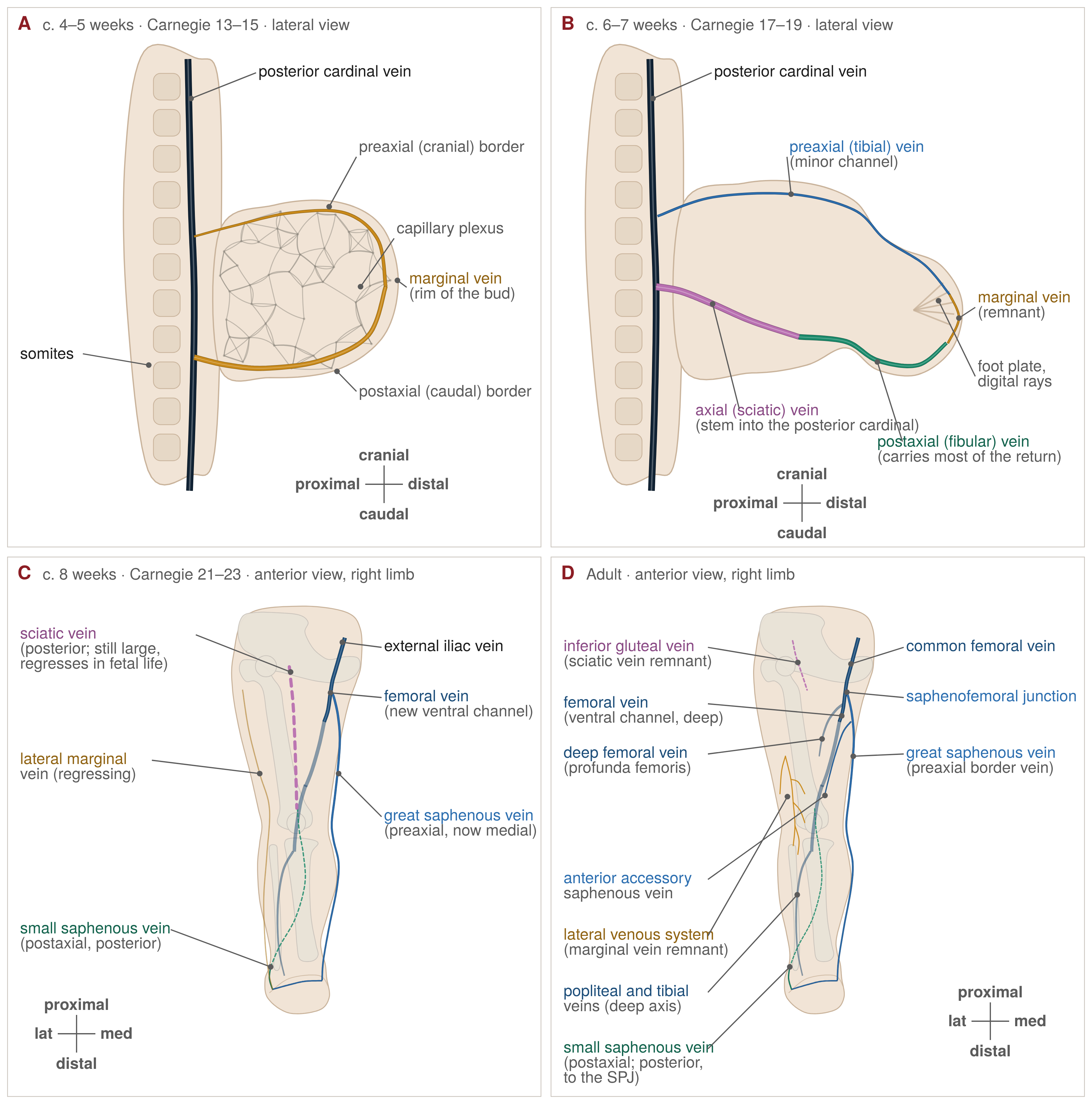
The veins of the leg are laid down in three overlapping phases: a capillary plexus drained by a single marginal vein, an axial phase in which a fibular channel and the sciatic vein carry the limb's blood to the pelvis, and a late preaxial phase in which the femoral and great saphenous veins take over and the earlier channels regress. Almost every variant the vein practitioner meets at the duplex machine — true saphenous duplication, the anterior accessory saphenous vein, an absent saphenopopliteal junction, the lateral venous system, duplicated femoral veins, persistent sciatic and marginal veins — is a recognisable fragment of that sequence. This review sets out what is known, what is inferred from animal embryos, and what it means for mapping, ablation and the management of congenital venous anomalies.
Why embryology matters at the duplex machine
No guideline asks the vein practitioner to know embryology. What the guidelines do ask is that treatment be planned on the patient's own "duplex anatomy", because the superficial veins are too variable for a textbook diagram.1 Embryology is the reason for that variability, and the framework that turns a list of variants into a pattern that can be anticipated.
The sonographer who knows that the great saphenous and anterior accessory saphenous veins are sister channels of one preaxial plexus will look for the second trunk when the first is found. The surgeon who knows that the femoral vein is a late arrival, replacing an older axial channel along the sciatic nerve, will not be surprised by a duplicated femoral vein or by a large popliteal-fossa vein that is not the popliteal. The practitioner who recognises a lateral marginal vein as a persistent embryonic trunk rather than a varicose tributary will prove the deep system before touching it. The 2002 nomenclature consensus already encodes some of this, defining the lateral venous system as the remnant of the embryonic lateral marginal vein and the sciatic vein as the main trunk of the primordial deep venous system.2
How we know: staging and the limits of the evidence
Human embryos are staged by the Carnegie system of O'Rahilly and Müller, by morphology rather than estimated age.3 Stage 13, at about 28 post-ovulatory days, is the first at which both limb buds can be seen; stage 15, at about 33 days, shows a lower limb bud with a tapering caudal half that will form the foot; by stage 17, at about 41 days, there is a digital plate.3 The stage descriptions say nothing about limb-bud veins. Uhl, whose work from the Paris computer-assisted anatomical dissection group is the fullest modern account, places the lower limb bud at about day 33, later than the Carnegie description, which is preferred here. On his framework organogenesis occupies weeks 4–8 (stages 13–23), the fetal period begins at week 9, and venous anatomy is adult-like by the end of week 12.3,4
The textbook account rests on early twentieth-century series: Hochstetter's description of 1893, the earliest mammalian account, and Lewis's rabbit embryos of 1905, from which the sequence of marginal, fibular, sciatic and femoral veins is taken.4,5 Woollard's three stages of limb vascular differentiation — capillary network, retiform plexus, definitive vessels — were described in the pig forelimb and reach most practitioners through the Hamburg classification built on them.6,7 Uhl is explicit that the data on the early stages "are from small mammals and need to be confirmed by direct observations in human embryos".4
The human evidence is thin. The only modern three-dimensional reconstruction of fetal limb veins, by Kurobe and colleagues, studied two fetuses at 14 and 15 weeks and found in every limb a large axial vein along the sciatic nerve while the femoral vein was still a small plexus.8 Two fetuses cannot define a timetable, but they show that the adult "persistent sciatic vein" is the normal configuration at the end of organogenesis and that the switch to a femoral-dominant system is a fetal event. Uhl's adult dissection material supplies the other end of the bracket: a modal femoral axis in 88% of adults, an axiofemoral bitruncular pattern in 9% and a single axiofemoral trunk in 3%.4 When the human sciatic vein regresses is therefore an inference, bracketed between "large at 14–15 weeks" and a small arcade in nine adults out of ten.
The developmental sequence
The capillary plexus and the marginal vein
The limb bud appears as a flat plate of mesenchyme supplied by a central axial artery and drained by an undifferentiated subcutaneous capillary network.4 At stage 16, about 37 days, Uhl describes the first "primitive veins" as endothelial migration thickens the distal and caudal edge of the plate into a marginal venous sinus.4 This marginal (border) vein runs around the free edge of the plate and drains, in Lewis's rabbits, into a primitive fibular vein, then an ischiadic (sciatic) vein and so into the posterior cardinal vein; Lee's formulation is the same.4,5,6
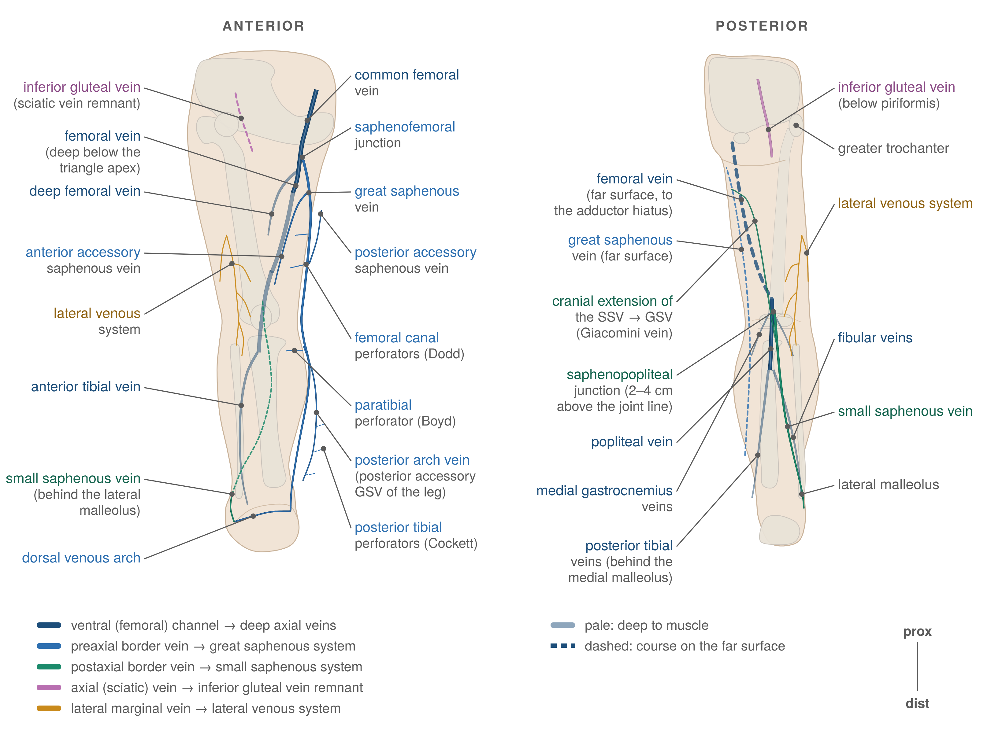
Two adult structures descend directly from this phase. The distal marginal vein, on Uhl's reading, becomes the primitive small saphenous vein, in his words the very first adult vein to appear in the embryo.4 Along the lateral leg a thin marginal network persists in most adults, connected to the fibular veins by perforators at fairly constant sites about 10, 14, 17 and 22 cm from the apex of the lateral malleolus.4 That network is the lateral subdermic venous system, which the 2002 consensus names the lateral venous system, the remnant of the embryonic vena marginalis lateralis.2
The postaxial and axial phase: fibular and sciatic veins
By day 17 in the rabbit the fibular vein is the main vein of the limb.5 It is fed from the sciatic vein above and, in Lee's account, branches below into the anterior tibial vein and a connecting branch, which together form the sciatic vein, "the second embryonic vein".6 Uhl places the primitive anterior tibial vein at about week 5 and the posterior tibial vein among the structures present by stage 23, about 56 days.4 The sciatic (axial, ischiadic) vein runs with the sciatic nerve, becomes large at weeks 7–8 and joins the deep femoral vein above; at that stage it is the main trunk of the thigh.4 Textbooks describe this phase as a postaxial (fibular) border vein, from which the small saphenous vein and the lateral marginal remnant descend, and a preaxial (tibial) border vein, from which the great saphenous vein descends; the modern sources describe the same relationships as caudal and cranial plexuses, and the preaxial/postaxial formulation is taken from the standard embryology texts rather than from a primary series.4,6
The preaxial takeover: the femoral vein
The decisive event for the adult deep system is a new ventral channel. In Lewis's rabbits an anastomosis from the sciatic vein connects to a new femoral vein, which becomes the definitive deep system while the sciatic vein regresses; Lee describes the same connecting branch, failure of the transition leaving the sciatic vein as the limb's main drainage.5,6 Uhl is more specific: the femoral vein develops along the femoral nerve from the cranial plexus, connects upward with the sciatic vein to form the iliac vein, and is at first small and plexiform; the anastomosis of this cranial plexus with the axial plexus gives rise to what he calls the "popliteal crossroad", the meeting point of so many variants.4
Uhl's central hypothesis is that three "angio-guiding nerves" — femoral, sciatic and the cutaneous saphenous nerves — appear at stage 19, about 47 days, and that the venous plexuses condense along them by stage 23. The proposed mechanism, VEGF from axons and Schwann cells, is extrapolated from arterial patterning in mouse skin, and Uhl presents it as a hypothesis not yet confirmed in embryos.4 It remains a useful mnemonic: each saphenous trunk, the sciatic vein and the femoral vein run with the nerve of the same name.
The saphenous veins, limb rotation and the saphenous compartment
The great saphenous vein appears with the femoral vein along the femoral nerve, from the cranial plexus.4 The caudal plexus forms the distal small saphenous vein together with a cranial extension in the thigh; a possible anastomosis with the axial plexus leads to termination in the popliteal axis, the saphenopopliteal junction.4 That is why the small saphenous vein so often has a thigh extension and why its popliteal junction is inconstant: the junction is a secondary anastomosis. Lee's account differs — the femoral vein continuing as the posterior tibial vein and serving as the precursor of the great saphenous vein — and the two have not been reconciled.4,6
Between weeks 6 and 8 the limb rotates medially, so that its cranial (preaxial) aspect faces medially and its caudal (postaxial) aspect laterally.4 That places the preaxial great saphenous vein medially and the postaxial small saphenous vein and marginal remnant laterally and posteriorly, which is why lateral-system reticular veins and spider veins cluster on the lateral thigh.
The saphenous trunks end up in a compartment of their own. Caggiati's study of 64 limbs showed a fibro-elastic lamina in the hypodermis of the medial thigh and leg and the back of the leg which, with the muscular fascia, forms compartments containing the saphenous veins and nerves; the trunks are therefore not truly "superficial", a term that fits only their tributaries.9 The developmental origin of the saphenous fascia has not been described, but the compartment, formalised by the 2002 consensus, is what distinguishes a trunk from a tributary on ultrasound.2
The cardinal systems, iliac veins and IVC
The limb veins drain into a trunk system that is itself being rebuilt. The paired posterior cardinal veins drain the caudal embryo; between weeks 4 and 8 three further paired systems appear — subcardinal, supracardinal and, in the Langman tradition, sacrocardinal.10 In the summary of Ghandour and colleagues, the posterior cardinals mostly regress, their distal portions becoming the iliac confluence and the common and external iliac veins; the right subcardinal forms the suprarenal IVC, the right supracardinal the infrarenal IVC, and the right vitelline vein the suprahepatic IVC.10 Whether the iliac veins derive from the caudal posterior cardinals or from separate sacrocardinal veins is a textbook-level disagreement, not a settled point.
The trunk veins begin bilaterally symmetrical and the left side regresses: double IVC, in about 2–3% of people, reflects persistence of both infrarenal supracardinals, and a left-sided IVC, in about 0.5%, caudal regression of the right supracardinal.7 These matter to the vein practitioner chiefly through iliofemoral thrombosis in the young.
Valves and perforators
Valves are a late, flow-dependent addition. Kampmeier and Birch, in the classical description of 1927 as summarised by Caggiati and by Bazigou and Makinen, described endothelial ridges invaded by mesenchyme to form the anlage, orientation toward the heart, widening of the cusps with enlargement of the sinus, and thinning of the sinus wall; valves appear only after the heart beats and the limb muscles move, the first in the lower limb at about 3–4 months in the deep veins of the femoral trigone and popliteal fossa and in the upper great saphenous vein.11 The Poznań fetal series supports this: in 110 fetal great saphenous veins the earliest well-formed valves appeared at 13 weeks, when each vein carried 2–7, and only bicuspid valves were seen.12 Valve number rises through prenatal life, some valves are lost after birth, and in the adult the terminal valve is present in 98–99% and the preterminal in 70–85%.13 In mice, femoral-vein valves form postnatally, after flow is established and the vein has acquired a smooth-muscle coat, at stereotyped positions downstream of tributary entrances; the timing does not transfer to humans but the sequence does.14
Perforating veins are the one element for which no timed developmental account could be found: Uhl's figures label femoral perforating branches and his text describes the fibular perforators, but no retrieved source says when or how they form.4 The 2002 consensus settles the vocabulary: perforating veins link the superficial and deep systems, communicating veins link veins within one system, and perforators are named by position, not eponym.2
| Embryonic channel | Adult derivative | Persists abnormally as |
|---|---|---|
| Marginal (border) vein of the limb plate | Lateral venous system of the thigh and leg; distal part becomes the small saphenous vein2,4 | Persistent lateral marginal vein (valveless, fascia-less trunk from lateral foot to thigh or pelvis)6 |
| Primitive fibular (postaxial) vein | Fibular veins and fibular perforators to the marginal remnant4 | Contributes to the lateral marginal vein when the second-stage transition fails6 |
| Sciatic (axial, ischiadic) vein | Small arcade along the sciatic nerve; collateral capacity for the femoral vein2,4 | Persistent sciatic vein (complete, upper or lower type), often with a hypoplastic femoral or popliteal vein6,8 |
| Cranial (preaxial) plexus along the femoral nerve | Femoral vein, great saphenous vein, anterior and posterior accessory saphenous veins4 | Duplicated or plexiform femoral vein; true great saphenous duplication; axiofemoral variants (12% of adults)4 |
| Caudal (postaxial) plexus | Small saphenous vein with cranial extension; secondary anastomosis forms the saphenopopliteal junction4 | Absent saphenopopliteal junction; small saphenous vein continuing as the Giacomini vein4 |
| Posterior cardinal veins (caudal part) | Iliac confluence, common and external iliac veins10 | Iliac aplasia or hypoplasia with collateral pathways10 |
| Subcardinal and supracardinal veins | Suprarenal and infrarenal IVC, left renal vein, azygos and hemiazygos10 | Double IVC (2–3%), left IVC (0.5%), azygos continuation, infrarenal agenesis7,10 |
Molecular control, briefly
The classical account is purely morphological: a plexus, selection of some channels, regression of others. Molecular work shows that arterial and venous identity are specified before blood flows: in the mouse, ephrin-B2 marks arterial and its receptor Eph-B4 venous endothelium in the primary plexus before circulation begins, and arterial fate is driven by Notch signalling downstream of VEGF, Dll4 initiating and Dll1 maintaining it.15 Venous identity is not a default: the nuclear receptor COUP-TFII is expressed in venous but not arterial endothelium, and its venous-specific deletion activates Notch so that veins acquire arterial features.16 Flow then remodels what the genetic programme has set.15
Venous valves borrow the lymphatic programme: valve-forming venous endothelium induces ephrin-B2 early, developing and mature valves express Prox1, Vegfr3 and integrin-α9, and ephrin-B2 and integrin-α9 signalling are required for formation and maintenance, loss of integrin-α9 in adult mice causing leaflet regression.14 Human relevance comes from mutation carriers: FOXC2 carriers had about 75% fewer venous valves than controls, with leaflets less than half normal length, and GJC2 (connexin-47) carriers about 62% fewer.17 Primary deep venous incompetence in a young patient with lymphoedema-distichiasis is valve morphogenesis failing.
None of this explains why the sciatic channel regresses and the femoral channel is selected; no retrieved study links a gene to either event. What molecular work does supply is a mechanism for malformation. Somatic TEK (TIE2) mutations were found in 28 of 57 (49.1%) sporadic venous malformations,18 and somatic hotspot PIK3CA mutations in 27 of 50 (54%) TEK-negative malformations, both producing chronic AKT activation on a common TIE2–PI3K–AKT pathway.19 A somatic PIK3CA C420R variant at an allele fraction of 4.7% has been found in an excised upper-limb marginal vein in CLOVES syndrome, and the authors argue for "anomalous marginal vein" rather than "persistent embryonic vein".20 Post-zygotic mosaic events in endothelium during the plexus period supply a basis for the Hamburg "extratruncular" lesion — arrested reticular tissue that retains proliferative capacity — and fit Oduber's hypothesis that the persistent embryonic veins of Klippel–Trénaunay syndrome arise from "a genetic defect leading to abnormal venous pattern formation".21 Whether a lower-limb persistent marginal or sciatic vein carries such a mutation has not been shown.
Variants the embryology explains
GSV duplication and the saphenous eye
Few variants have been reported across so wide a range as "double" great saphenous vein: about 1% to nearly 50% by method and definition.22 Kockaert and colleagues showed the range to be a definition problem. True duplication is two trunks both lying between the fascial blades of the saphenous compartment, each with its own "eye"; partial duplication is a trunk not fully enclosed; closed loops are partial duplications that reconverge at or above the knee; and parallel or "false" veins are channels outside the fascia, most often the anterior accessory saphenous vein, usually smaller and draining a different territory.22 In their retrospective duplex series of 240 examinations true duplication was present in 4 (1.6%) and partial in 59 (24.6%); in a prospective series of 100 untreated patients, 2% and 17%.22 Phlebography cannot image fascia and so cannot make the distinction, which is why the older phlebographic rates are high. The UIP duplex consensus gives true duplication — two parallel veins in the same compartment for 3–25 cm of thigh — as under 1%, with the eye sign the key to separating a true trunk from a parallel tributary.23
A true duplication is a second intrafascial channel surviving from the plexiform cranial plexus; a "false" duplication is a sister vein that never entered the compartment. Kockaert and colleagues recommend treating a confirmed duplication, suggest that a missed second trunk may partly explain recurrence, and note that no data show whether a competent second trunk refluxes after ablation of the first.22
Accessory saphenous veins
The anterior accessory saphenous vein is present in 41% of subjects, typically joining within 1 cm of the saphenofemoral junction, and is involved in about 14% of patients with varicose veins.23 The 2002 consensus defines the anterior, posterior and superficial accessory veins by their position relative to the great saphenous vein and its fascia.2 The junction itself is inconstant: a meta-analysis of 16 studies and 7,433 legs found a bifid junction in 9.6% and two separate junctions in 1.7%, with four tributaries the commonest number — the authors' reason for recommending pre-operative duplex.24
The accessory veins are preaxial channels of the same plexus as the trunk, and the 2024 multisociety series on the "anterior saphenous vein" draws the clinical conclusion: on ultrasound of 62 normal and 62 varicose limbs the vein lies in its own compartment and "must be considered a truncal vein", treated like the great or small saphenous vein rather than as a tributary.25 One-year results of the SYNCHRONOUS study, reported at Charing Cross in 2024 in 1,173 patients, showed that after great saphenous ablation an untreated competent anterior accessory vein over 2 mm developed reflux in 13.7%, one under 2 mm in 4.8%, against 0.9% when ablated at the same sitting; these are conference data awaiting publication.26 The SVS/AVF/AVLS guideline of 2022 suggests endovenous ablation, with phlebectomy if needed, over ligation and stripping for symptomatic anterior accessory reflux (Grade 2, Quality C), and includes that vein and the small saphenous vein in the standard reflux examination.27
SSV termination and the cranial extension
The saphenopopliteal junction is a secondary anastomosis between the caudal and axial plexuses, and its variability follows.4 In the dissection series of Uhl, Gillot and Lemasle, a junction less than 5 cm above the crease was present in 83% (62% without and 21% with a common trunk with the medial gastrocnemius veins), a high junction in 6%, no junction with continuation as the Giacomini vein in 5%, plexiform termination in the thigh muscles in 5%, and a short termination at leg level in 1%; in 30% the vein ended low on the popliteal vein, crossing the medial side of the tibial nerve.28 A duplex study of 244 limbs in young volunteers with a meta-analysis of 13 studies gave quite different pooled figures: no true junction, the vein continuing as a prominent thigh extension, in 24.4% (95% CI 19.3–29.5); termination in the popliteal vein without any cranial extension in 17.1%; the other patterns combined in 54.7%.29 The UIP consensus places the junction most often 2–4 cm above the crease, higher in 25%, with gastrocnemius veins joining the small saphenous vein first in 10–30% of limbs.23
A thigh extension is the rule — 95% of limbs by the UIP figure — and the 2002 consensus names it the cranial extension of the small saphenous vein, "often termed" the vein of Giacomini when it joins the great saphenous vein via the posterior thigh circumflex vein.2,23 Delis and colleagues reported a single termination in 86.8% and dual in 12.7%, the great saphenous vein the commonest cranial endpoint (49.5%), and reflux in the Giacomini vein in 3.3% of 301 limbs with suspected venous disease.30
The nerves travel with the vein because they guided it. In 20 cadaver legs, Kerver and colleagues found multiple junctions in 20%, a mean vein-to-tibial-nerve distance of 4.4 mm (under 5 mm in 55% of legs, 1 mm or less in 20%), and the sural nerve within 5 mm of the vein in 60% at the tibial plateau, rising to 100% from 200 mm distally.31 Whoever treats this vein by radiofrequency ablation is working in the embryonic nerve–vein corridor.
The lateral subdermic venous system
Albanese and colleagues described the lateral subdermic venous system in 1969 as a distinct network on the lateral thigh and leg and the usual source of lateral-thigh reticular veins and telangiectasia, a description that reaches most readers through later texts.32 The 2002 consensus adopted "lateral venous system" with the embryological definition already quoted,2 and Uhl's fibular perforators explain why lateral telangiectasia often has a reticular feeder communicating with the fibular veins rather than the saphenous trunks.4 No retrieved study gives a population prevalence or sex ratio.
Deep-vein duplication
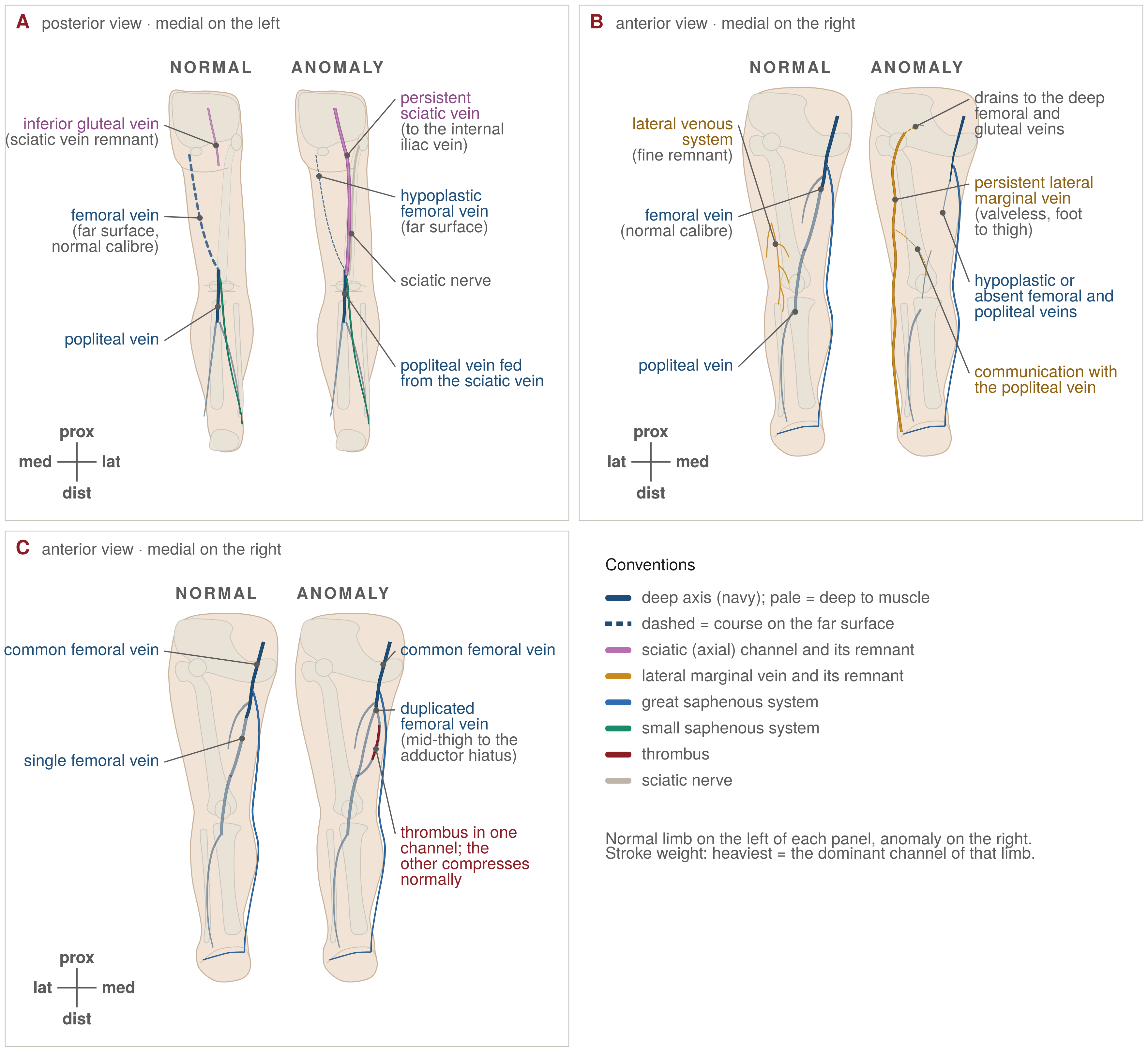
Because the femoral vein condenses late from a plexus, incomplete coalescence leaves parallel trunks. In 404 bilateral venograms from a thromboprophylaxis trial, Quinlan and colleagues found a duplicated femoral vein in 31% of 808 limbs and a true duplicated popliteal vein in 5%, half the duplications beginning in the mid-thigh, with strong bilateral concordance.33 Screaton and colleagues found multiple femoral veins in 46% of 381 venograms after a negative ultrasound for suspected thrombosis.34 Park and colleagues, with bilateral CT venography in 445 patients, found femoral or popliteal multiplication in 29% of limbs, a course variation relative to the artery in 8%, and agenesis of both femoral and popliteal veins, with a persistent sciatic vein as the sole axis, in 0.3% of limbs and 0.7% of patients.35 A meta-analysis of 11 studies and 3,682 limbs put pooled femoral duplication at 19.7% (95% CI 11–30), with a wide gap between cadaver series (2%) and imaging (25%), presumably because collapsed duplicates are missed at dissection.36
| Variant | Reported prevalence | Population / method |
|---|---|---|
| True GSV duplication (both trunks intrafascial) | 1.6% and 2%; <1% | Duplex, 240 retrospective and 100 prospective examinations;22 UIP consensus23 |
| Partial GSV duplication / closed loops | 24.6% and 17% | Same duplex series22 |
| Anterior accessory saphenous vein present | 41%; pathological in ~14% of varicose limbs | UIP consensus23 |
| Bifid SFJ / two separate junctions | 9.6% / 1.7% | Meta-analysis, 16 studies, 7,433 legs24 |
| SSV thigh (cranial) extension | 95% | UIP consensus23 |
| No SPJ, SSV continuing as a thigh extension (Giacomini pattern) | 5%; 24.4% (95% CI 19.3–29.5) | Dissection;28 duplex in 244 limbs plus meta-analysis of 13 studies29 |
| High SPJ (≥5 cm above crease) | 6% | Dissection28 |
| Multiple SPJs | 20% | 20 cadaver legs31 |
| Duplicated femoral vein | 31%; 46%; 19.7% pooled (cadaver 2%, imaging 25%) | 808 venographic limbs;33 381 venograms after negative ultrasound;34 meta-analysis, 3,682 limbs36 |
| True duplicated popliteal vein | 5% | 808 venographic limbs33 |
| Femoral or popliteal multiplication | 29% of limbs | Bilateral CT venography, 445 patients35 |
| Axiofemoral (non-modal) femoral axis | 12% (bitruncular 9%, unitruncular 3%) | Adult dissection material4 |
| Femoral and popliteal agenesis (persistent sciatic vein as sole axis) | 0.3% of limbs; 0.7% of patients | Bilateral CT venography35 |
| Double IVC / left IVC | 2–3% / 0.5% | SVS/AVF haemodynamics chapter7 |
When embryonic channels persist
Persistent sciatic vein
The persistent sciatic vein is the embryonic axial vein that was never superseded. Cherry, Gloviczki and Stanson, as summarised in the recent anatomical literature, divided it into a complete type from the popliteal vein through the sciatic notch to the internal iliac vein, an upper type from upper-thigh tributaries ending in the pelvis, and a lower type confined to the thigh and draining into the deep femoral vein or a subcutaneous plexus; they found 20 among 279 patients with Klippel–Trénaunay syndrome.37 Reported prevalence ranges from 0.7% to 9% across cadaver and phlebographic series, figures the reviewers regard as uncertain; magnetic resonance venography is the most reliable test.37 Koç and colleagues described bilateral lower-type veins with hypoplastic popliteal veins in a 93-year-old cadaver, each sciatic vein carrying the main drainage and containing well-formed valves.38 The 2002 consensus retains the term sciatic vein and notes its capacity as a collateral for the femoral vein.2
The series of Labropoulos and colleagues, 24 limbs with reflux in veins along the sciatic nerve, is a selected cohort without a denominator, but it carries the warning that matters: one patient with acute thrombosis of a persistent sciatic vein died of pulmonary embolism. Nineteen limbs had pain or tingling in the nerve distribution; subfascial ligation, phlebectomy or sclerotherapy relieved or improved 17.39 A persistent sciatic vein lies in the plane of a popliteal sciatic nerve block, and a lower-type vein with a hypoplastic popliteal vein may be the limb's main outflow — an argument against ligating it during popliteal surgery and against saphenectomy when the sciatic channel is the drainage.37,38
Persistent lateral marginal vein and Klippel–Trénaunay
Lee's position, carried in the title of his 2014 paper, is that the marginal vein "is not a varicose vein; it is a venous malformation": a truncular lesion in the Hamburg sense, valveless, with a smooth-muscle-deficient wall and no fascial sheath, running from the lateral foot to the thigh, buttock and sometimes the internal iliac vein, the commonest venous malformation in Klippel–Trénaunay syndrome.40 Its frequency in that syndrome varies with the cohort and the definition: 12 of 70 (17.1%) in the Dutch duplex series of Oduber and colleagues, under 20% in the review by Fereydooni and Nassiri as cited by Gloviczki, and 72% for varicosities or venous malformations of any kind, including atypical lateral and sciatic veins, among 252 Mayo Clinic patients.21,41,42,43 Oduber and colleagues reduce the many names for persistent embryonic veins to two entities, persistent lateral marginal vein and persistent sciatic vein, each reported with a statement of deep venous patency.21
Deep venous patency is the whole question: deep aplasia or hypoplasia accompanies the marginal vein in 13% of cases in the Fereydooni and Nassiri review and in "more than one third" in Lee's experience.40,41 Lee's principles follow: duplex is the test of choice, because phlebography alone can falsely suggest aplasia; excision is the treatment when the deep system is normal; with mild hypoplasia, staged removal under perioperative low-molecular-weight heparin; with aplasia the marginal vein "is part of the main drainage, so resection is not possible".40 Rojas Martinez and colleagues make the same point — if the deep system is aplastic or hypoplastic, extensive varicectomy "should not be performed, because these veins act as substitute channels" — and add that the commonest diagnostic error is to mistake a marginal vein for an arteriovenous malformation, which must be excluded.44 The Mayo series of open surgery in 49 patients (53 limbs; lateral embryonic vein excised in 28%) recorded deep-vein thrombosis in 4%, pulmonary embolism in 2% and peroneal palsy in 2%, with freedom from reintervention of 78% at three years.45 The marginal vein is an embolic source: 94% of patients with the syndrome who had a pulmonary embolism had ectatic change in the marginal vein, as cited by Gloviczki.41,42
IVC and iliac anomalies
The practitioner who scans for deep-vein thrombosis meets the cardinal systems as IVC agenesis. Population incidence is under 0.01%, but about 5% of patients under 30 with idiopathic thrombosis, particularly men, have an IVC anomaly; bilateral iliofemoral thrombosis occurs in under 10% of thromboses overall but in up to 50% with an IVC malformation; and most authors favour long-term anticoagulation on low-level evidence — figures cited from the review by Gómez-Hernández and colleagues.46 Sun and colleagues, in 64 patients with congenital aplasia, atresia or hypoplasia of the iliac and lower-limb veins, found a left-sided and male predominance, multi-segment defects in 58% and deep-vein aplasia in 12%, and described five collateral pathways that must not be mistaken for pathological vessels.47 Iliac compression at the arterial crossing is best regarded as an acquired lesion on a developmentally determined crossing, not a malformation.
The Hamburg/ISSVA view of malformations
The Hamburg classification groups congenital vascular malformations by predominant vessel and splits each into extratruncular lesions — arrested before the truncal stage, mesodermal remnants that may proliferate after trauma, surgery or hormonal change — and truncular lesions (aplasia, hypoplasia, stenosis, aneurysm, persistent embryonic vein), which do not proliferate but have worse haemodynamic consequences.6 Uhl ties the stages to time: malformations before week 4 are extratruncular and severe, those of weeks 4–8 truncular and less severe, and after week 8 only simple variants arise.4 The ISSVA classification adds a section for malformations of named vessels (the former "truncular" category), lists the known genes, and defines Klippel–Trénaunay syndrome as a capillary–lymphatic–venous malformation with limb overgrowth, Parkes Weber as capillary malformation with arteriovenous fistulae and overgrowth, and CLOVES as lymphatic–venous–capillary malformation with lipomatous overgrowth.48 The VASCERN-VASCA working group adds management pathways for sporadic, multifocal, familial and syndromic venous malformations.49 Hamburg says how a lesion will behave; ISSVA what to call it and which gene to seek.
| Clinical consequence | Reported rate | Source population |
|---|---|---|
| False-negative compression ultrasound for proximal DVT with a duplicated femoral vein | 6% (10/177) vs 2% (4/204) with a single vein, P = 0.056 | 381 venograms after negative ultrasound34 |
| Pulmonary embolism from an ectatic marginal vein in KTS | 94% of KTS patients with PE had ectatic change in the marginal vein | Review, as cited by Gloviczki41,42 |
| DVT and PE after open venous surgery in KTS | DVT 4%, PE 2%, peroneal palsy 2% | 49 patients, 53 limbs, Mayo Clinic45 |
| Fatal PE from persistent sciatic vein thrombosis | 1 death in 24 limbs | Selected clinical series39 |
| Nerve injury with small saphenous vein treatment | 3.0% pooled, all modalities | Network meta-analysis, 6,793 limbs50 |
| Tibial nerve within 5 mm of the SSV near the junction | 55% of legs; within 1 mm in 20% | 20 cadaver legs31 |
| Reflux developing in an untreated competent AASV after GSV ablation | 13.7% (>2 mm), 4.8% (<2 mm) vs 0.9% if ablated synchronously, at 1 year | 1,173 patients, conference report26 |
| Absent saphenopopliteal junction (SSV continues as a thigh extension) | 5%; 24.4% pooled | Dissection;28 duplex plus meta-analysis29 |
| Deep venous aplasia or hypoplasia accompanying a marginal vein | 13%; "more than one third" | Review, as cited by Gloviczki;41,42 Lee's experience40 |
| IVC anomaly in idiopathic DVT under 30 years | ~5%; bilateral iliofemoral DVT up to 50% with IVC malformation | As cited by Gómez-Hernández46 |
Work-up before intervention
Every variant in this review is detectable on a complete duplex examination performed with the question in mind, which is why the author maps every limb fully before offering any treatment, whether radiofrequency ablation, medical adhesive closure or ultrasound-guided foam sclerotherapy. The ESVS guideline describes duplex as the primary test, performed upright, the deep veins checked for patency, reflux and post-thrombotic obstruction, and the findings drawn as a venous map, "an essential tool to plan and perform venous interventions".1 Where a marginal or persistent sciatic vein is suspected, duplex is the first test but not the last: Lee advises adding venography, and plethysmography helps where the marginal vein's contribution to drainage is in doubt.40,42
Before you treat…
- Count the trunks in the compartment. Two intrafascial channels, each with its own eye, are a true duplication; a channel outside the fascia is an accessory vein or tributary.22,23
- Find the anterior accessory saphenous vein and measure it. It joins within about 1 cm of the junction and runs over the femoral vessels; a competent vein over 2 mm is at risk after great saphenous ablation.23,26
- Map the junction tributaries. A bifid junction or two separate junctions is not rare.24
- Follow the small saphenous vein to its end. Is there a junction at all, how high, do gastrocnemius veins share a trunk, and does a cranial extension carry flow — in which direction?1,28,29
- Identify the tibial and sural nerves before choosing the access point.31
- Look for the lateral system when lateral-thigh reticular veins or telangiectasia are the complaint, and trace the feeder to its perforator.1,4
- Prove deep patency and count the deep channels. Compress each separately; if one limb has a duplication, look carefully at the other.33,34,36
- A large vein beside the sciatic nerve or along the lateral limb changes the plan. Establish the deep system, exclude arteriovenous shunting, and consider cross-sectional venography before any intervention.21,40,44
What the guidelines say
Guideline positions on duplex mapping, variants and congenital anomalies
ESVS 2022. Duplex ultrasound is the primary diagnostic test of choice, and because of the "large variability" of the superficial veins it is "mandatory to rely on the so called 'duplex anatomy' to plan any treatment". The anterior accessory saphenous vein runs parallel and slightly lateral to the great saphenous vein in its own compartment; the small saphenous junction level "can vary"; and "In the presence of a Giacomini vein, the saphenopopliteal junction may be present or absent." For small saphenous incompetence, endovenous thermal ablation is recommended over surgery or foam (Class I, Level A), with care to avoid sural nerve injury if cannulation is below mid-calf (Class I, Level B); for reticular veins and telangiectasia, duplex is recommended to look for associated incompetent veins, which are treated first (Class I, Level C). The guideline "decided to leave out congenital venous malformations", so it contains no recommendation on the marginal vein or Klippel–Trénaunay syndrome.1
SVS/AVF/AVLS 2022 (Part I). Duplex is "the diagnostic test of choice to evaluate for venous reflux" (Grade 1B); reflux is more than 500 ms in superficial truncal, tibial, deep femoral and perforating veins and more than 1 s in the common femoral, femoral and popliteal veins; the standard examination includes spectral Doppler of the great saphenous vein, "the anterior accessory great saphenous vein and … the small saphenous vein", with diameters measured in each. Guideline 2.2.3 suggests endovenous ablation over ligation and stripping for symptomatic anterior accessory reflux (Grade 2C). The text does not discuss great saphenous duplication or the saphenous eye, and does not address congenital anomalies.27
UIP duplex consensus 2006 (Part II, Anatomy). The anatomical reference for the saphenous eye, the 41% prevalence of the anterior accessory vein, true duplication under 1%, saphenopopliteal variability and the 95% thigh extension.23
Nomenclature consensus 2002. The source of the terms used here, including the embryological definitions of the lateral venous system and the sciatic vein.2
VASCERN-VASCA and ISSVA. Cover the syndromic malformations; no document specific to the marginal vein was identified, and the most explicit management statements remain those of Lee, of Fereydooni and Nassiri, and of the Mayo group.40,41,42,48,49
Where the guidelines are silent. None addresses embryology as such, true saphenous duplication, the persistent sciatic vein, or the timing of accessory-vein treatment relative to great saphenous ablation beyond the single SVS suggestion above.
Practical implications for treatment
The findings that change what is done at the time of treatment are collected in Table 4. The author offers radiofrequency ablation, medical adhesive closure and ultrasound-guided foam sclerotherapy, not endovenous laser or mechanochemical ablation; comments on those modalities are drawn from the literature cited.
| Finding | Best evidence | Practical points |
|---|---|---|
| True GSV duplication | Duplex definition series; no outcome data on the fate of a competent second trunk22 | Treat a refluxing duplicate as a trunk; document a competent duplicate and follow it. Each trunk needs its own access and its own tumescence or adhesive run. |
| Anterior accessory saphenous vein at the time of GSV treatment | Conference data from 1,173 patients; SVS Grade 2C for ablation of symptomatic reflux26,27 | Treat it as a truncal vein. Ablate a refluxing accessory vein at the same sitting; discuss synchronous treatment of a competent vein over 2 mm, acknowledging that the supporting data are not yet fully published. |
| SSV access and termination | Cadaver nerve-distance data; ESVS Class I B on cannulation level; pooled nerve injury 3.0%1,31,50 | Cannulate at mid-calf; stop the catheter short of the junction with the tibial nerve in view; where there is no junction, treat the vein as a Giacomini continuation and define its outflow before ablating. |
| Giacomini vein | Small treatment series; one common peroneal injury after thermal ablation, as reviewed by Nowak-Tarnawska and Reina-Gutierrez30,51 | Establish direction of flow; the posterior thigh course lies close to nerve; foam or adhesive avoid thermal injury but neither has been compared with ablation in this vein. |
| Lateral venous system | ESVS Class I C: duplex for associated incompetence before treating telangiectasia1 | Treat the reticular feeder, and any fibular perforator feeding it, before the telangiectasia; foam sclerotherapy is the usual tool for the feeder. |
| Duplicated femoral or popliteal vein | Venographic and CT series; false-negative ultrasound 6% vs 2%33,34,35 | Compress each channel separately when excluding thrombosis; a duplication is a reason to doubt a single negative scan in a patient with a high pre-test probability. |
| Persistent sciatic vein | Case series and cadaver reports37,38,39 | Never ablate or ligate the only outflow. With a hypoplastic popliteal or femoral vein the sciatic vein is the drainage; symptomatic reflux in a sciatic-nerve vein has been treated by subfascial ligation or sclerotherapy only after deep patency was confirmed. |
| Lateral marginal vein | Expert series; Mayo surgical outcomes; management reviews40,41,42,44,45 | Prove deep patency and exclude arteriovenous shunting first. With a normal deep system, excision is the treatment of choice; with hypoplasia, staged removal under LMWH; with aplasia, do not remove. Ligate or coil the perforators before any foam so sclerosant cannot enter the deep system; thermal ablation is generally unsuitable under thin skin and foam is less effective and carries risk; give weight-adjusted LMWH around any intervention. |
| IVC or iliac anomaly | Case series; low-level evidence on anticoagulation46,47 | Image the IVC in a young patient with unexplained or bilateral iliofemoral thrombosis; recognise collateral pathways as compensatory and do not treat them as varicose veins. |
A working algorithm
- Perform a complete bilateral duplex map, standing, including deep-vein patency and channel count, both saphenous compartments, the anterior accessory saphenous vein, the small saphenous termination and the lateral thigh.1,27
- If a large valveless vein runs along the lateral limb or beside the sciatic nerve, classify it as a persistent lateral marginal or persistent sciatic vein, state the deep venous patency, exclude arteriovenous shunting, and obtain cross-sectional venography before planning anything.21,40,44
- If the deep system is aplastic, the embryonic vein is the drainage: compression, thromboprophylaxis and referral to a vascular-anomalies centre, not ablation.40,42
- If the deep system is patent, treat truncal reflux by the modality suited to the vein — the great saphenous vein and a true duplicate, the anterior accessory saphenous vein and the small saphenous vein each as trunks, the accessory vein addressed at the same sitting when it refluxes.25,27
- Treat reticular feeders and perforators of the lateral system before telangiectasia, and tributaries after the trunk.1
- Document every variant on the map and in the letter: the next clinician to scan the limb for suspected thrombosis needs to know about the second femoral channel.33,34
Areas of uncertainty
- The human timetable is inferred. The sciatic-to-femoral switch comes from rabbit embryos; the only modern human series is two fetuses.4,5,8
- The origin of the great saphenous vein is described three ways — preaxial border vein, cranial plexus along the femoral nerve, or outgrowth of the femoral–posterior tibial line — and the accounts have not been reconciled.4,6
- The "angio-guiding nerve" hypothesis is unproven in embryos, and perforator development has not been described.4
- The molecular account is mouse-derived. Human confirmation rests on 13-week fetal valves and mutation-carrier phenotypes; no gene has been linked to the selection or regression of a limb channel, and no somatic mutation has been shown in a lower-limb persistent sciatic or marginal vein.12,14,15,17,20
- Prevalence depends on method. Femoral duplication is 2% at dissection and 25% on imaging; the saphenopopliteal junction is absent in 5% or 24% depending on the series.28,29,36
- Prophylactic treatment of a competent duplicate or accessory trunk rests on one unpublished trial and expert opinion; the guidelines are silent on embryology, true duplication, the persistent sciatic vein and the marginal vein.1,22,26,27
Key clinical points
- The leg veins develop in three phases — marginal, axial (sciatic) and preaxial (femoral) — and the clinically important variants are fragments of the earlier phases.4,6
- The human evidence is thin: the sequence rests on rabbit embryos and two human fetuses, and the timings here are approximate.5,8
- The saphenous compartment defines a trunk. True great saphenous duplication occurs in about 1–2%; most "double" veins are accessory veins or loops.22,23
- The anterior accessory saphenous vein is a sister trunk, present in about 41% and pathological in about 14% of varicose limbs; a competent vein over 2 mm left at great saphenous ablation refluxed in 13.7% at one year in conference data.23,26
- The saphenopopliteal junction is a secondary anastomosis, absent in 5–24% of limbs; follow the small saphenous vein to its true outflow and identify the tibial and sural nerves before access.28,29,31
- The lateral venous system is the marginal-vein remnant, with fibular perforators; map the feeder before treating lateral-thigh telangiectasia.2,4
- The femoral vein is a late channel, duplicated in about a fifth to a third of limbs on imaging; compress each channel when excluding thrombosis.33,34,36
- A large vein beside the sciatic nerve may be the limb's drainage. Never ligate or ablate it without proving the femoral and popliteal veins.37,38
- The marginal vein is a venous malformation, not a varicose vein. Deep aplasia accompanies it in 13% to more than a third; prove the deep system and exclude arteriovenous shunting before any treatment.40,41,44
- Guidelines require treatment to be planned on the patient's duplex anatomy, but are silent on embryology and on persistent embryonic veins.1,27
A bedside rule
Before ablating any vein, name the embryonic channel it came from, and never close a channel until you have seen the one that replaced it.
Disclosure. The author performs endovenous and other venous procedures in private practice. This article is for medical education and does not replace individual clinical assessment.
Figures. Diagrams drawn for this review by Vein Centres South Africa.
How to cite
Blignaut J. Embryology of the leg veins: a practical guide for the vein practitioner. Clinical review. Vein Centres South Africa; October 2026. Available from: https://www.veinsurgery.co.za/for-doctors/leg-venous-embryology/
References
- De Maeseneer MG, Kakkos SK, Aherne T, et al. European Society for Vascular Surgery (ESVS) 2022 clinical practice guidelines on the management of chronic venous disease of the lower limbs. Eur J Vasc Endovasc Surg 2022;63:184-267.
- Caggiati A, Bergan JJ, Gloviczki P, et al. Nomenclature of the veins of the lower limbs: an international interdisciplinary consensus statement. J Vasc Surg 2002;36:416-422.
- O'Rahilly R, Müller F. Developmental stages in human embryos. Washington, DC: Carnegie Institution of Washington; 1987. Publication 637.
- Uhl JF. Focus on venous embryogenesis of the human lower limbs. Phlebolymphology 2015;22:55-63.
- Lewis FT. The development of the veins in the limbs of rabbit embryos. Am J Anat 1905;5:113-120.
- Lee BB. Venous embryology: the key to understanding anomalous venous conditions. Phlebolymphology 2012;19:170-181.
- Meissner MH, Moneta G, Burnand K, et al. The hemodynamics and diagnosis of venous disease. J Vasc Surg 2007;46 Suppl:4S-24S.
- Kurobe N, Hakkakian L, Chahim M, et al. Three-dimensional reconstruction of the lower limb's venous system in human fetuses using the computer-assisted anatomical dissection (CAAD) technique. Surg Radiol Anat 2015;37:231-238.
- Caggiati A. The saphenous venous compartments. Surg Radiol Anat 1999;21:29-34.
- Ghandour A, Partovi S, Karuppasamy K, et al. Congenital anomalies of the IVC—embryological perspectives and clinical relevance. Cardiovasc Diagn Ther 2016;6:482-492.
- Kampmeier OF, Birch CLF. The origin and development of the venous valves, with particular reference to the saphenous district. Am J Anat 1927;38:451-499.
- Czarniawska-Grzesińska M, Bruska M. The structure of the cusps of valves in the human foetal great saphenous vein. Folia Morphol (Warsz) 2003;62:275-276.
- Caggiati A. The venous valves of the lower limbs. Phlebolymphology 2013;20:87-95.
- Bazigou E, Makinen T. Flow control in our vessels: vascular valves make sure there is no way back. Cell Mol Life Sci 2013;70:1055-1066.
- Kume T. Specification of arterial, venous, and lymphatic endothelial cells during embryonic development. Histol Histopathol 2010;25:637-646.
- You LR, Lin FJ, Lee CT, et al. Suppression of Notch signalling by the COUP-TFII transcription factor regulates vein identity. Nature 2005;435:98-104.
- Lyons O, Saha P, Seet C, et al. Human venous valve disease caused by mutations in FOXC2 and GJC2. J Exp Med 2017;214:2437-2452.
- Limaye N, Wouters V, Uebelhoer M, et al. Somatic mutations in angiopoietin receptor gene TEK cause solitary and multiple sporadic venous malformations. Nat Genet 2009;41:118-124.
- Limaye N, Kangas J, Mendola A, et al. Somatic activating PIK3CA mutations cause venous malformation. Am J Hum Genet 2015;97:914-921.
- Lim Y, Fereydooni A, Brahmandam A, et al. Mechanochemical and surgical ablation of an anomalous upper extremity marginal vein in CLOVES syndrome identifies PIK3CA as the culprit gene mutation. J Vasc Surg Cases Innov Tech 2020;6:438-442.
- Oduber CEU, Young-Afat DA, van der Wal AC, et al. The persistent embryonic vein in Klippel-Trenaunay syndrome. Vasc Med 2013;18:185-191.
- Kockaert M, de Roos KP, van Dijk L, et al. Duplication of the great saphenous vein: a definition problem and implications for therapy. Dermatol Surg 2012;38:77-82.
- Cavezzi A, Labropoulos N, Partsch H, et al. Duplex ultrasound investigation of the veins in chronic venous disease of the lower limbs — UIP consensus document. Part II. Anatomy. Eur J Vasc Endovasc Surg 2006;31:288-299.
- Cirocchi R, Henry BM, Rambotti M, et al. Systematic review and meta-analysis of the anatomic variants of the saphenofemoral junction. J Vasc Surg Venous Lymphat Disord 2019;7:128-138.e7.
- Caggiati A, Labropoulos N, Boyle EM, et al. The anterior saphenous vein. Part 2. Anatomic considerations in normal and refluxing patients. J Vasc Surg Venous Lymphat Disord 2024;12:101855.
- Venous News. One-year results for synchronous anterior accessory saphenous vein ablation hold up [Charing Cross Symposium 2024 report]. 2024. Available from: https://venousnews.com/one-year-results-for-synchronous-anterior-accessory-saphenous-vein-ablation-hold-up/
- Gloviczki P, Lawrence PF, Wasan SM, et al. The 2022 Society for Vascular Surgery, American Venous Forum, and American Vein and Lymphatic Society clinical practice guidelines for the management of varicose veins of the lower extremities. Part I. Duplex scanning and treatment of superficial truncal reflux. J Vasc Surg Venous Lymphat Disord 2023;11:231-261.e6.
- Uhl JF, Gillot C, Lemasle P. Relationship between the small saphenous vein and nerves: implications for the management of chronic venous disease. Phlebolymphology 2006;13:22-27.
- Veselá M, Beneš M, Dostálová G, et al. Variations of the saphenopopliteal junction: an ultrasonography study in a young population, a systematic review and a meta-analysis. Angiology 2024;75:527-535.
- Delis KT, Knaggs AL, Khodabakhsh P. Prevalence, anatomic patterns, valvular competence, and clinical significance of the Giacomini vein. J Vasc Surg 2004;40:1174-1183.
- Kerver ALA, van der Ham AC, Theeuwes HP, et al. The surgical anatomy of the small saphenous vein and adjacent nerves in relation to endovenous thermal ablation. J Vasc Surg 2012;56:181-188.
- Albanese AR, Albanese AM, Albanese EF. The lateral subdermic venous system of the legs. Vasc Surg 1969;3:81-89.
- Quinlan DJ, Alikhan R, Gishen P, et al. Variations in lower limb venous anatomy: implications for US diagnosis of deep vein thrombosis. Radiology 2003;228:443-448.
- Screaton NJ, Gillard JH, Berman LH, et al. Duplicated superficial femoral veins: a source of error in the sonographic investigation of deep vein thrombosis. Radiology 1998;206:397-401.
- Park EA, Chung JW, Lee W, et al. Three-dimensional evaluation of the anatomic variations of the femoral vein and popliteal vein in relation to the accompanying artery by using CT venography. Korean J Radiol 2011;12:327-340.
- Sibuor W, Kipkorir V, Cheruiyot I, et al. Prevalence of femoral vein duplication: systematic review and meta-analysis. J Ultrason 2021;21:e326-e331.
- Büyükertan M, Söztanacı US, Balcıoğlu HA. Persistent sciatic vein. Folia Morphol (Warsz) 2022;81:1062-1065.
- Koç T, Gilan İY, Külekçi GD, et al. Bilateral persistent sciatic vein: report of a case with developmental, histological and clinical aspects. Surg Radiol Anat 2014;36:189-194.
- Labropoulos N, Tassiopoulos AK, Gasparis AP, et al. Veins along the course of the sciatic nerve. J Vasc Surg 2009;49:690-696.
- Lee BB. Marginal vein is not a varicose vein; it is a venous malformation. Veins and Lymphatics 2014;3:4050.
- Fereydooni A, Nassiri N. Evaluation and management of the lateral marginal vein in Klippel-Trénaunay and other PIK3CA-related overgrowth syndromes. J Vasc Surg Venous Lymphat Disord 2020;8:482-493.
- Gloviczki P. Treatment and pitfalls: lateral marginal vein [conference slides]. VEITHsymposium 2024; abstract 827. Available from: https://veithsymposium.org/2024/abstracts/vei/827.pdf
- Jacob AG, Driscoll DJ, Shaughnessy WJ, et al. Klippel-Trénaunay syndrome: spectrum and management. Mayo Clin Proc 1998;73:28-36.
- Rojas Martinez R, Puech-Leão P, Guimarães PM, et al. Persistence of the embryonic lateral marginal vein: report of two cases. Rev Hosp Clin Fac Med S Paulo 2001;56:159-162.
- Malgor RD, Gloviczki P, Fahrni J, et al. Surgical treatment of varicose veins and venous malformations in Klippel-Trenaunay syndrome. Phlebology 2016;31:209-215.
- Gómez-Hernández C, Fuentes García MI, Hernández-García C. Intrahepatic agenesis of the vena cava causing venous thrombosis of the lower limbs. Emergencias 2021;33:158-160.
- Sun P, Wang H, Xu W, et al. Anatomical patterns and collateral pathways in congenital aplasia, atresia, and hypoplasia of iliac and lower extremity veins: a retrospective cohort study. Surg Radiol Anat 2026;48:30.
- Wassef M, Blei F, Adams D, et al. Vascular anomalies classification: recommendations from the International Society for the Study of Vascular Anomalies. Pediatrics 2015;136:e203-e214.
- Dompmartin A, Baselga E, Boon LM, et al. The VASCERN-VASCA Working Group diagnostic and management pathways for venous malformations. J Vasc Anom 2023;4:e064.
- Oud S, Alozai T, Jamaludin FS, et al. Anatomic success of treatment modalities for small saphenous vein incompetence: a systematic review and network meta-analysis. Eur J Vasc Endovasc Surg 2025 (in press).
- Nowak-Tarnawska A, Reina-Gutierrez L. The vein of Giacomini: an in-depth review of its anatomy, hemodynamics, and treatment alternatives. Phlebolymphology 2024;31(1).