
About Spider Veins
Spider veins are small blue or red veins located just below the outer surface of the skin. They often have a web-like appearance, hence, the term spider vein. Most spider veins have an unattractive cosmetic effect and, in some cases, may give rise to symptoms such as itching, burning, or throbbing of the legs. Veins are arranged in layers within and under the skin. The very smallest type of veins are on the surface, these veins drain into slightly larger veins, called reticular veins, which lie deeper within the skin. Faulty reticular veins often act as "feeding" veins to the smaller superficial spider veins. The most prominent reticular vein is the lateral vein of the thigh. This vein is often involved in causing spider veins of the outside thigh. Treating the smaller veins on the surface, without addressing this important vein, leads to poor treatment results.

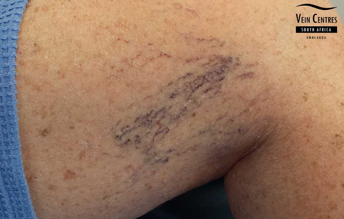
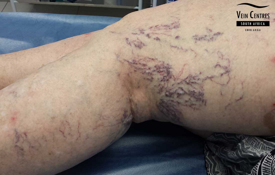

Spider Veins
What are spider veins?
Spider veins are like varicose veins but much smaller. They are thin red or blue lines, close to the surface of the skin. They can look like tree branches or spiderwebs with their short, jagged lines. Spider veins are found on the legs and face and can cover either a small or very large area of skin.
Spider veins symptoms
Most spider veins do not cause any symptoms apart from the cosmetic issues. Some patients will report burning or itching in their larger spider veins. These symptoms are often worse on warm days or with monthly menses in women. The actual vein, in a spider vein, is translucent, the colour that is observed depends on the colour of the blood flowing through the vein. Very small superficial spider veins are red, and can often look like a bruise. Larger spider veins, located deeper within the skin, often have a blueish appearance. Patients with spider veins who also have significant leg swelling, heaviness, pain or skin changes may have underlying venous reflux. In these patients, duplex ultrasound can determine whether an underlying venous problem should be addressed before cosmetic treatment1. Not sure whether your spider veins are only cosmetic? Read Spider veins: cosmetic or medical?
Spider veins causes
Spider veins are common and affect 50-80% of the general population, both men and women. All skin types are prone to the development of spider veins, for obvious reasons they are more visible in light skin and less obvious in darker skin. A family history of vein disease in parents or siblings predict a higher risk of developing spider veins. Other causes like trauma to the skin accounts for a small portion of spider veins.
Myths
There are many myths around the causes of spider veins today. Some of these include crossing your legs, wearing high heels, taking very hot baths and wearing tight clothes.
Using venotonic drugs and creams will remove my spider veins. There are a variety of online and printed ads promoting creams and medicines to cure your veins. There is no good evidence that creams or oral preparations remove spider veins.
Spider veins treatment
Sclerotherapy is an established first-line treatment for many lower-limb spider and reticular veins12. The most appropriate treatment depends on the size, depth and distribution of the veins and whether underlying reflux is present. Sclerotherapy is the treatment we use for spider veins at our centres. Over many years of treating vein patients, we have developed our treatment protocols. This means we use drugs indicated for the treatment of veins. We do not use solutions of salt or sugar water in treating veins, as practiced in the past. The dose of sclerosant each patient receives is carefully controlled, which helps limit side effects such as pigmentation and matting3. Compression stockings are worn after treatment, as compression has been shown to reduce pigmentation and matting following sclerotherapy.
New spider veins can form over time after treatment, and further sessions may be needed. Many patients see a good cosmetic improvement that lasts for years, while others develop new veins sooner. Individual factors such as family history, pregnancy and hormone therapy influence this. * Individual results may vary.
Spider vein treatment is therefore best thought of as ongoing maintenance rather than a once-off procedure, and top-up sessions are common.
Alternative treatments
Several alternative treatments are marketed for spider veins. The evidence supporting them varies considerably, and patients sometimes come to us disappointed with the results of treatments tried elsewhere.
Tablets and dietary supplements for veins
Many tablets and supplements are sold with the promise to keep your legs healthy and improve the symptoms of vein disease. These drugs are known as venotonic drugs and often based on red vine leaf extract. Unfortunately the scientific evidence used to market these drugs is limited at best. I always find it interesting that a patient would choose to take these drugs for many years without having the root cause treated. The long term effects of these drugs are not known. In our practice we have had very few patients actually reporting these drugs to be useful. There might be a role for venotonic drugs in patients who suffer from post thrombotic syndrome.
Creams
Urea creams, Vitamin K creams the list goes on. At best these creams might have a temporary effect at hiding the veins, but certainly there is no proof of treatment. Some creams are intended to mask the veins, similar to using make-up.
Laser or IPL
Some clinics use laser or intense pulsed light (IPL) devices to treat leg spider veins. These devices operate at a wavelength that targets haemoglobin, the red pigment of blood, and they are highly effective for the fine red vessels of the face and neck.
Randomised comparisons of long-pulsed Nd:YAG laser against sclerotherapy for leg telangiectasias have found broadly comparable clearance by the end of follow up, with sclerotherapy producing improvement more quickly and laser treatment reported as more painful45. A network meta-analysis published in 2023 found Nd:YAG laser performed well for the smallest telangiectasias6. It is worth noting what those trials studied: patients selected by specialists, with isolated surface veins and no untreated underlying reflux. They do not tell us how laser performs when it is used without a full assessment of the leg.
That distinction matters, because in practice laser is often used on leg veins without a duplex scan having been done. In our experience laser gives good results in straightforward cases where the spider veins are truly isolated, but where reticular feeder veins are involved the results are poor and we see early recurrence. Surface light-based treatment acts only on the vessel visible at the skin surface, whereas the feeder veins that supply it sit deeper in the skin.
Our practice follows the old phlebology rule of thumb: laser above the level of the heart, sclerotherapy below it. For the face and neck, laser is excellent. For the legs, we use sclerotherapy, which allows the reticular feeder veins to be treated in the same session. Every patient has a duplex scan first, so that any underlying reflux is identified and addressed before surface veins are treated.
Laser treatment also carries a risk of skin burns, since enough heat energy has to reach the vein to destroy it. Burnt skin heals over time but can be left de-pigmented (whiter than the surrounding skin) or pink, shiny and hyperpigmented (darker than the surrounding skin). This risk is greater in darker skin types.
Spider veins prevention
Our current understanding of spider veins would suggest that you cannot prevent spider veins from forming.
Spider Veins Diagnosis
Venous duplex ultrasound is the investigation of choice for diagnosing vein disease1. It involves the use of an ultrasound machine and high frequency probe to accurately assess the anatomy of your veins as well as the flow patterns within them. Ultrasound scanners do not produce any radiation (unlike x-rays) and they don’t involve needles or injections. Hand-held dopplers are not used in our vein centres as they are inaccurate. Every new patient will undergo a duplex examination prior to any treatment planning. Treating surface veins without identifying any underlying venous problem is more likely to give a disappointing result. This is often why we see patients who have been treated elsewhere presenting with a worsening in their veins.
Spider Veins and Sclerotherapy
References
- De Maeseneer MG, Kakkos SK, Aherne T, et al. European Society for Vascular Surgery (ESVS) 2022 clinical practice guidelines on the management of chronic venous disease of the lower limbs. Eur J Vasc Endovasc Surg. 2022;63(2):184–267. ESVS guideline
- Gloviczki P, Lawrence PF, Wasan SM, et al. The 2022 SVS, AVF and AVLS clinical practice guidelines for the management of varicose veins of the lower extremities. Part I. J Vasc Surg Venous Lymphat Disord. 2023;11(2):231–61. Journal
- Goldman MP, Sadick NS, Weiss RA. Cutaneous necrosis, telangiectatic matting, and hyperpigmentation following sclerotherapy: etiology, prevention, and treatment. Dermatol Surg. 1995;21(1):19–29. PubMed 7600016
- Parlar B, Blazek C, Cazzaniga S, et al. Treatment of lower extremity telangiectasias in women by foam sclerotherapy vs. Nd:YAG laser: a prospective, comparative, randomized, open-label trial. J Eur Acad Dermatol Venereol. 2015;29(3):549–54. PubMed 25069999
- Munia MA, Wolosker N, Munia CG, Chao WS, Puech-Leão P. Comparison of laser versus sclerotherapy in the treatment of lower extremity telangiectases: a prospective study. Dermatol Surg. 2012;38(4):635–9. PubMed 22221551
- Bontinis V, Bontinis A, Koutsoumpelis A, et al. Interventions for the treatment of lower limb telangiectasias and reticular veins: a systematic review and network meta-analysis. Eur J Vasc Endovasc Surg. 2023. EJVES
Written and medically reviewed by Dr Johan Blignaut, MBChB (UOFS), FCS(SA)SURG, Specialist Surgeon.
Last medically reviewed: August 2026. This page is reviewed periodically and updated when clinical practice changes. It is general information, not medical advice – see our terms and medical disclaimer.